Dr. Gus A. Wright | Core Facility Manager & Assistant Research Scientist
Email: gwright@cvm.tamu.edu | Tel: 979-458-9859
Office: VMR Addition 274 | Core Facilities: VMR 210, VMA 214D & VMA 214C
Please contact Dr. Wright before using any of the core equipment for the first time to schedule a training session.
There is no scheduling of equipment—it’s first come, first use!
Dr. Wright provides assistance in experimental design, use of equipment, and analysis of microscopy images, upon request.
Core Equipment
Microscopes: Brightfield, Fluorescence, and Stereo
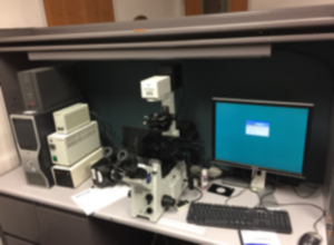
Olympus IX 71 Inverted Microscope with SPOT RT Slider Camera
Brightfiedl/DIC/Fluorescence (DAPI, FITC, Texas Red)
VMR 210
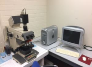
Olympus Vanox AH2 Upright Microscope with SPOT Insight Camera
Brightfield only
VMA 214D
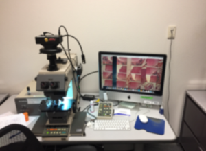
Olympus Vanox AH3 Upright Microscope with SPOT RT3 Slider Camera
Brightfield/DIC/Fluoresence (DAPI, FITC, Texas Red)
VMR 210
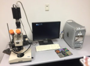
Olympus Research Stereo SZH Microscope with SPOT Insight Camera
Brightfield only
VMR 210
Gel Documentation Systems
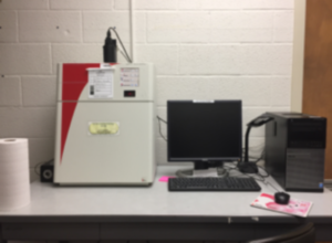
Alpha Innotech Fluorchem System
Primarily used for EtBr DNA gels and Coomassie stained protein gels
VMA 214C
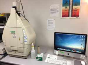
Biorad Chemidoc MP Gel Documentation System
DNA gels (EtBr, PI, sytox green, etc.), protein gels (Biorad stain free, coomassie, Ponceau Red, etc.)
Chemiluminescence, Flurescence (Blue LED, Green LED, Red LED)
VMR 210
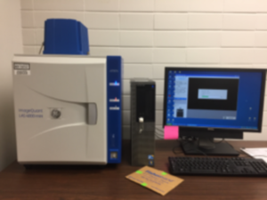
GE ImageQuant LAS 4000 Mini Chemiluminescence Gel Documtnation System
Chemiluminescence only
VMA 214C
Fluorescence Microplate Reader
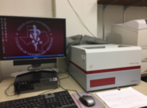
BMG Labtech Fluorstar Omega Fluorescence/Luminescence/Absorbance Microplate Reader
The FLUOstar Omega is a versatile, multi-mode microplate reader for five detection modes: fluorescence intensity (including FRET),
luminescence (including BRET), UV/Vis absorbance, time-resolved fluorescence (including TR-FRET), and AlphaScreen®/AlphaLISA®.
Uses MARS data analysis program.
VMR 210
Other Imaging Equipment
Nikon Coolscan 5000 for scanning 1”x 3” microscope slides
VMR 210