
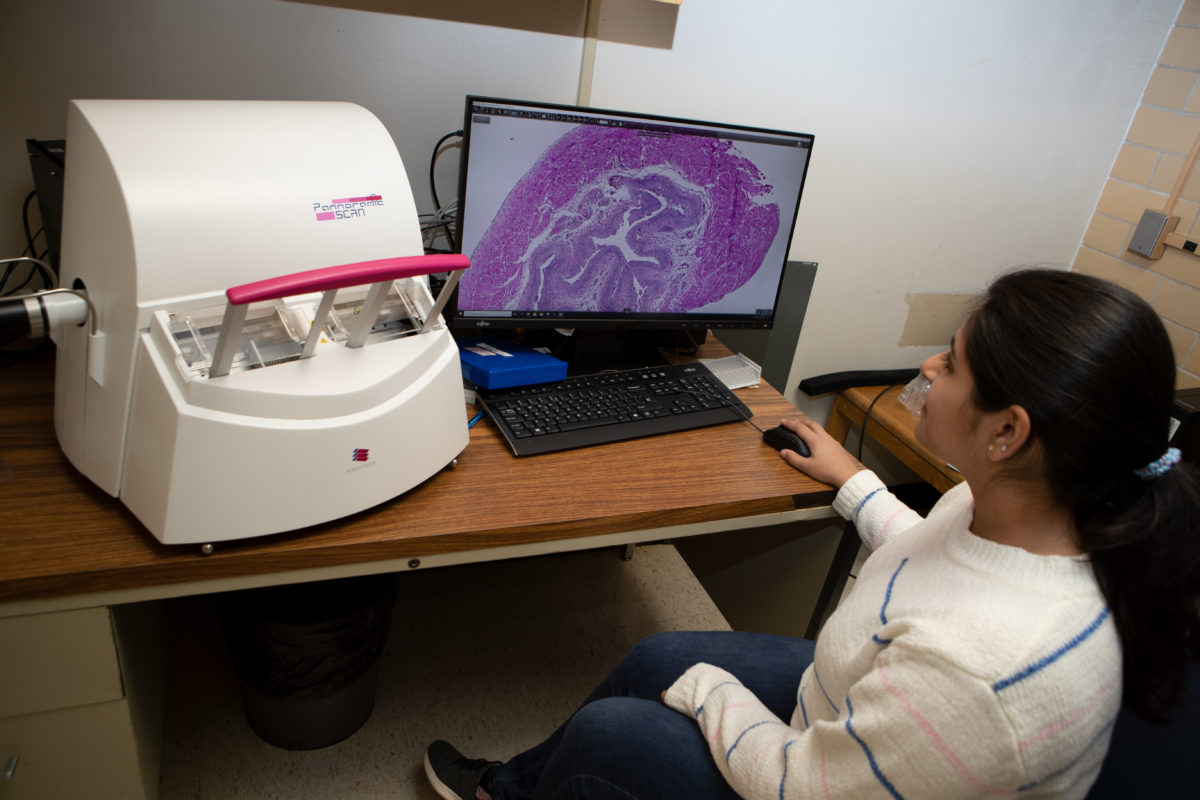
The VMBS Core Histology Laboratory offers digitization of histological slides using a state-of-the-art, whole slide scanner (Pannoramic Scan II made by 3DHistech®). The scanner produces best-in-class, digital Brightfield (BF) or Fluorescent (FL) images by a single layer or multi-layer (up to 30 levels) scanning at 20x or 40x objective.
The Pannoramic SCAN II supports the best Carl Zeiss objectives, achieving up to ~ 50x and 98.5x native optical resolution (scanned at 20x or 40x magnification, respectively). The system provides superb images that can be used (approved) for research and teaching.
**Note: For most histological evaluations, scanning at 20x is recommended and expected to be suitable.**
**Note: Scanned slides will be purged from our repository 30 days after the images are supplied to you. You will need to download them and save them in a personal location. Once purged, the images are not recoverable and slides will have to res-canned (at your expense) if you need access to them again.**
**Note: Slides that have an adhesive (“sticker”) label cannot be scanned as the label will potentially cause a malfunction with the scanner. If your slides have a sticker label you will need to remove it and any residual glue PRIOR to submission. Slides that are submitted with a sticker label will be returned to you without being scanned.
Scanning Services
- Digitization of whole slides at 20x or 40x magnification in BF or FL modes
- Single layered or multi-layered (up to 30 layers)
- Extended autofocus for both 20x and 40x scans
- FL scanning in up to 6 different fluorescent channels (DAPI/FITC/TRITC/Cy5/Cy7/Texas Red)
- NOTE: FL images can currently only be viewed using Case Viewer, on a PC platform. Images cannot be processed on a Mac platform (the download available for Mac computers that is on the 3D Histech site does not work with our system).
- Image management using the Hamamatsu Server which allows for viewing and management of BF images on a PC or Mac
- The slide viewing software (Slide Viewer) is free to download and install on your PC computer to view both BF and FL images
- SlideViewer opens several 3rd party digital slides: SVS files (Aperio by Leica Biosystems) and NDP, VMS, VMU files (Hamamatsu) and supports multiple file export formats: TIFF, JPEG, SVS, MRXS.
- Slides will be delivered and stored in SVS format as the default. If you prefer mrxs format, please indicate so at the time of submission.
- **Comparison of images in both formats has been done and quality is not lost with the SVS conversion.
Submission of scan requests:
- Submission of scan requests for research is done online and for teaching is done via submission of the excel form below:
- Online submission: You must submit your requests for RESEARCH scans using this portal.
- Submission form for teaching
- Complete the form and print it out to bring with your slides or email the completed form to digitalpathology@cvm.tamu.edu.
- A member of the research histology unit will contact you to arrange a drop-off time and discuss the delivery of your images after your slides are scanned.
- Form must be received in order for the lab to take possession of your slides.
- Allow for a minimum of 2 weeks for completion of your scans.
Pathology Services
Dr. Jones-Hall is a board-certified veterinary pathologist who specializes in experimental pathology. She is skilled at histological and pathological evaluation of all organ systems of vertebrate animals and can evaluate multiple stains including immunohistochemistry and cytochemistry. She uses morphometry to better assess disease, or the lack thereof. She uses an advanced Zeiss microscope and its software to evaluate and photograph tissues via traditional light microscopy. She utilizes Visiopharm® analytical software for computer-augmented, quantifiable, and precise, high-throughput evaluations of pathology in tissue.
Dr. Jones-Hall is available for:
- Collaboration; preliminary consultations; or hourly, fee-based analyses
- Assistance with the pathology-related verbiage for grants, manuscripts, and presentations (on projects that she has worked on)
- Graduate committee membership, if desired, for student projects where she has made a significant contribution
Questions? Concerns? Email digitalpathology@cvm.tamu.edu.