
Leading in Animal Oncology
Veterinarians at the Texas A&M College of Veterinary Medicine & Biomedical Sciences (CVM) serve on the front lines of the war on cancer. Some of their discoveries not only save beloved pets’ lives but also offer hope for treating cancer in humans. As the fastest growing disease on earth, cancer has been the focus of a national “war” proclaimed by every United States president since Richard M. Nixon. By 2030, experts believe there will be 22 million cases of cancer in humans worldwide.
A Longtime Leader in Animal Oncology
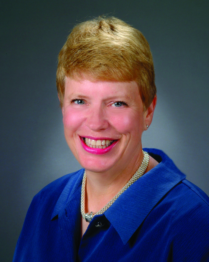
As a pioneer in animal oncology, Dr. Claudia Barton, a CVM professor in the Department of Small Animal Clinical Sciences (VSCS), has seen the field go through monumental changes during the past 40 years. Soon after her arrival at Texas A&M; University in 1976, Barton remembers a retiring veterinarian handing her multiple boxes of Kodachromes, or slides, of cancer tumors. “The only treatment that anybody had done at that time on animals with cancer was surgery. That was about all there was,” she said.
Barton’s interest in oncology began when she earned her doctorate in veterinary medicine from the University of Missouri (UM). After graduation, Barton followed Dr. George Shelton, a former UM dean, who became the CVM dean in 1973. “We worked together when I was a student at Missouri, so he asked me if I wanted to pursue a residency in pathology at Texas A&M;,” Barton said. “I came here, did the residency, and then went back to teach at the University of Missouri. Then it was Easter Sunday, and I could not get my car out of the driveway because there was so much snow. A position came open at Texas A&M;, and the bluebonnets were blooming there, so I applied for it. I took a salary cut to come here, but I did not want to live in the snow.”
The field of animal oncology was quickly emerging when Barton returned to the CVM. She didn’t waste any time. Immediately, she began working with other top researchers to found the Veterinary Cancer Society. “We all got together in a little office around a table and just said, ‘We need to start a specialty of oncology,'” Barton said. “It was a dynamic group of people, including pathologists, surgeons, and internal medicine folks, who were interested in cancer.” Today, the professional society serves more than 800 members internationally.
Early Advances in Radiation Therapy and Chemotherapy
The CVM was among the first colleges to offer radiation therapy for animals. Initially, the veterinarians used an orthovoltage unit, which was a routinely used diagnostic radiology machine. A human hospital eventually donated the more advanced cobalt teletherapy unit to the CVM. “It was difficult and expensive to actually bring this unit into the radiation department because they had to build a shielded facility with many layers of concrete in the walls so that the radiation would not permeate outside of the room,” Barton said.
Although these early radiation technologies were helpful for diagnosing and treating cancer, they did pose a risk to the animal. “The problem with this type of therapy was that you had to use a high dose to get any penetration into deep tissues, so there were a lot of radiation burns,” Barton said.
During these early years of treatment options and while chemotherapy drugs were still in their infancy, CVM researchers found radiation therapy to be a useful procedure when surgery wasn’t an option. “As chemotherapy advanced, a few drugs were developed, but our veterinarians didn’t have access to them,” Barton said. “Most of the drugs that we use today became available for veterinary medical use after I began my career in oncology.”
The Advancement of Cancer Treatment in Animals
The introduction of imaging technologies, such as computed tomography (CT) scans, magnetic resonance imaging (MRI) scans, and positron emission tomography–computed tomography (PET-CT) scans, has moved veterinary medicine light-years ahead in the fight against cancer. “In the old days, if a dog came in with nasal bleeding and was found to have a nasal tumor on X-rays and radiographs, the only way surgeons could determine how extensive it was was to go in and start probing around to find out if it had gone behind the eye or broken through the bone between the nasal cavity and the brain,” Barton said. “Now, with CTs and MRIs, we don’t have to put dogs through those procedures, and it has been such an advance in veterinary medicine.”
These various scans provide important information to veterinarians as they diagnose and treat cancer. For instance, CVM veterinarians can use CT scans to combine X-ray images taken from different angles and then create cross-sectional images of bones, blood vessels, and soft tissues of an animal’s body through computer analysis. An MRI scan uses a strong magnetic field and radio waves to create detailed images of the animal’s organs and tissues. PET-CT scans, the latest imaging technology, allow researchers and clinicians to view any abnormal activity in the animal’s tissues and organs. This scan also can identify a target for biopsy and help researchers analyze the effectiveness of cancer treatments.
Barton believes advancing technology will open the way to new discoveries and better treatments. “We’ll be able to see, finally, tiny little foci of tumor cells that we can’t appreciate now because they are just too small to visualize,” she said. “I think we’ll even become more sophisticated in our ability to see where tumors are located and then to make the decision about whether you want to put an animal through treatment. Maybe you’re not going to want to put your animal through radiation therapy for its nasal tumor if you find out that it is already somewhere else. In the old days, we wouldn’t have known until they showed up with the clinical signs that the cancer has spread.”
Today’s CVM veterinarians are also more sophisticated in their knowledge of the different types of cancer. “It’s amazing to me that we used to make sweeping generalizations about cancers that were good and cancers that were bad with so little knowledge,” Barton said. “Every day, we advance our knowledge about the biologic behavior of these tumors related to molecular diagnostics that we had no idea about. What we do regularly today would have been considered something akin to Star Wars in those days. Our molecular diagnostic tests also have revolutionized our ability to give people prognoses for their animal’s cancer.”
Searching for Cancer Treatments for Both Dogs and Humans
Some CVM research discoveries also hold promise for helping humans who are diagnosed with cancer. For instance, the Texas Neuro-Oncology Program, which started in 2008, focuses on understanding deadly brain tumors. This partnership between the CVM, the University of Texas Medical School at Houston, and M.D. Anderson Cancer Center began when Dr. Stephen Fletcher, a UTMS pediatric neurosurgeon, saw his two boxers die of brain tumors. He reached out to the CVM’s Dr. Jonathan Levine, an associate professor, department head, and McWhorter Chair in VSCS. Their collaborative studies have found that the growth of spontaneous and highly aggressive brain tumors, such as glioblastoma multiforme in dogs, closely mirrors what happens in humans. The researchers hope to discover novel therapies that will work in dogs, with the hope that these therapies can be translated into a useful form for treating children with similar types of brain cancer.
Researchers also study human cancers to identify potential treatments for animals. When faced with an animal that has an unusual form of cancer, Barton and other CVM research faculty and veterinarians often delve into the National Cancer Institute’s database. “I’ll first start by going to cancer.gov, and I will think, ‘Okay, if this dog with this stage of this cancer were a human, what would they be doing for that human?'” Barton said. “Can we do that? Or is it too painful? Too difficult? Too technically beyond our means?”
Choosing Quality over Quantity
While CVM veterinarians and researchers strive to find new treatments, they also realize that some animals are not good candidates for therapy. Therefore, they try to help pet owners think through the ramifications and consequences of their healthcare decisions. “One of the things that we always have to think about is that the dog or cat or horse is not choosing this for themselves,” Barton said. “They don’t wake up in the morning and say, ‘I have to make it to Christmas. I have to make it to my child’s college graduation.’ When a dog wakes up in the morning, it thinks, ‘Is today a good day? Do I feel good? Do I get to play with my ball? Do I look forward to my dinner?’ When we take away their good days because of treatment when they’ve only got three months to live, are you going to take a month of that and make the animal not feel good?”
Ultimately, Barton believes it’s important to counsel the owner about the realities of cancer. “Of course, we’re trying to sort out for them what they really want for their animal, what’s right for their animal, and what we can and cannot accomplish,” she said. “You have to keep reminding them that many of these cancers do not have a cure. We’re trying to prolong the animal’s life with quality. We do not want quantity without quality.”
Oncology in both humans and animals has made tremendous strides over the past 40 years, and more breakthroughs are on the horizon. Barton believes that the future of cancer treatments in humans-and eventually animals-will be personalized medicine. “It will be beyond my practice career and probably beyond my life, but they will biopsy a human tumor, and they will find the cancer genome. And then, they will figure out what kinds of targeted molecular therapies will be available based on the genetics,” she explained. “They will apply that therapy, and then the cancer mutates and they will look at the genome again and figure out the next therapy. This process, ideally, allows you to turn cancer into a chronic disease.”

