
Blood Gases
Gas exchange occurs through the medium of blood. Blood circulates through the body, exchanging gases with tissues, and circulates in the lungs, exchanging gases with the air. Only two gases are important, oxygen and carbon dioxide. Do you remember from Cells Are Us why this is so? If not, review the unit on Energy. Why do we not care about nitrogen, which makes up 78% of the air? Nitrogen does not normally exchange in the sense that we discuss oxygen and carbon dioxide. Nitrogen only becomes important in scuba diving, where it bubbles out of the tissues if you have been deep in the water. How Gas Amount Changes| Gas | Lungs | Tissues |
|---|---|---|
| Blood oxygen | increases | decreases |
| Blood Carbon Dioxide | decreases | increases |
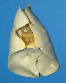
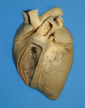 Cow heart, with part of the wall cut away to show interior of a pump chamber (ventricle). Note the valve at the top of the chamber. When blood fills the artery (part of its wall is also cut away) during contraction of the ventricle, the backpressure closes the valve.
Gas Exchange in Lungs
Oxygen & Carbon Dioxide The body requires oxygen in order to produce energy for our cells to do work. Therefore, it is essential that we have an efficient system of obtaining oxygen from the air. Our bodies also produce a toxic gas called carbon dioxide that must be efficiently removed from the body to prevent cell damage. When we inhale, we pick up oxygen from air. When we exhale, we flush out carbon dioxide. Each breath lasts only a few seconds (even less if we are running.) Isn’t it amazing that gas exchange occurs so quickly? What is it about gases that lets them exchange so quickly? The Structures of the Respiratory System Here is a list of structures to become familiar with:- Mouth and Nose- these are the openings where respiratory gases enter and leave the body.
- Trachea (windpipe)- this passage way connects the mouth and nose to the lungs.
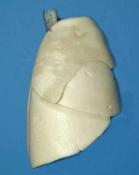
- Lungs- these are the balloon-like structures that temporarily hold air in the body.
- Bronchial tube- the trachea breaks up into these smaller tubes to enter the right and left lungs.
- Bronchioles- within the lungs the bronchi split into these even smaller tubes which attach to the alveoli.
- Alveoli- these are the small sac-like structures where gas exchange occurs with the blood.
- Air enters your body through your mouth and nose. The nasal passage connects to the oral passage at the back of the mouth where a tube called the trachea connects the mouth and nose to the lungs. Dirt from the air is filtered before it ever reaches the lungs by the hairs and mucus in the nose. This hair and mucus trap some of the dirt and germs found in the air to protect the lungs from infection.
-
- The trachea, or windpipe, is a tube that connects the nose and mouth to the lungs. You can see and feel your trachea on the front of your neck. The tube has C-shaped cartilaginous rings wrapped around it to prevent the tube from collapsing and blocking the air flow to and from the lungs. Air is the only substance designed to go down the trachea.Why do you think the trachea is only meant for air? What happens if something blocks the trachea?
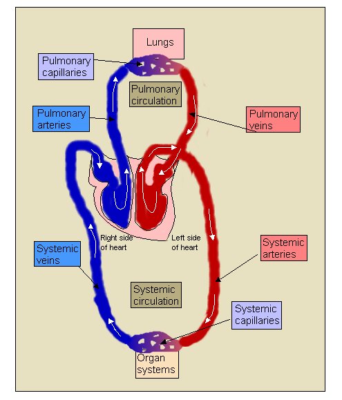
The System for Transporting Gases
Your heart beats with about the strength it takes to squeeze a tennis ball. Squeeze a tennis ball and see how hard that is. Now think what it must be like for your heart to do this 70 times a minute, 60 minutes an hour, 24 hours a day – for a lifetime!The Circulatory System
The heart, the blood, and the blood vessels make up a system for the transport of gases, nutrients, and chemical wastes. The primary functions of the circulatory system are the following:- To transport nutrients and oxygen to the cells.
- To remove waste and carbon dioxide from the cells.
- To provide for efficient gas exchange.
The Heart
The heart is made up of cardiac muscle and is divided into 4 different chambers. The top two compartments are called atria, while the bottom two compartments are called ventricles.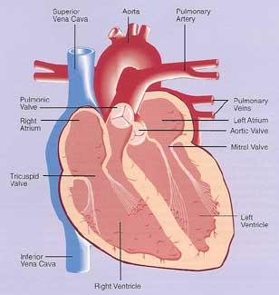
Blood Vessels
There are three basic types of blood vessels:- Arteries- these carry “oxygen rich” blood away from the heart, except in the case of the artery to the lungs.
- Capillaries- these are the sites of gas exchange between the tissues.
- Veins- these return “oxygen poor” blood to the heart, except for the vein that carries blood from the lungs.
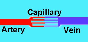 On the right is a diagram showing how the three connect. Notice the artery and vein are much larger than the capillaries. This junction is called a capillary bed.
The capillaries have very thin walls and there are many of them.
Why do you think this is?
Why are the capillaries shown with two different colors?
Why is the vein shown as blue?
On the right is a diagram showing how the three connect. Notice the artery and vein are much larger than the capillaries. This junction is called a capillary bed.
The capillaries have very thin walls and there are many of them.
Why do you think this is?
Why are the capillaries shown with two different colors?
Why is the vein shown as blue?
Alveoli
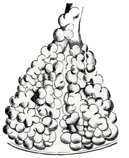 Exterior view of Alveoli. This is where gas exchange takes place in the lungs.
Alveoli are tiny balloon-like sacs, in the lungs, where gas exchange takes place and they serve as the barrier between the external environment (the air) and the internal environment (the blood).
Because gas must exchange in a second or two between blood vessels and the air inside of our lungs, how do you suppose the process is made efficient?
Exterior view of Alveoli. This is where gas exchange takes place in the lungs.
Alveoli are tiny balloon-like sacs, in the lungs, where gas exchange takes place and they serve as the barrier between the external environment (the air) and the internal environment (the blood).
Because gas must exchange in a second or two between blood vessels and the air inside of our lungs, how do you suppose the process is made efficient?
-
-
-
- These small sacs have very thin walls that are full of blood vessels called capillaries. The walls are so thin that the oxygen, brought in during inhalation, can diffuse through them to enter your blood.
- Likewise, carbon dioxide is carried by your blood to the blood vessels in the alveoli where it diffuses through the thin walls and into the air in your lungs. That “used” air is now ready for exhalation. Each lung has millions of alveoli, and your body needs all of them to get enough oxygen into your blood!
-
-
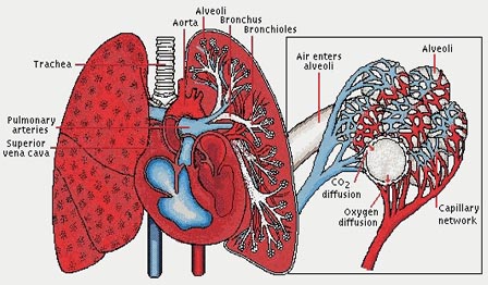 Why are there so many capillaries at the alveoli? Why are they shown in blue and red?
What is the problem with the buildup of tar and other hazardous materials from smoking in the lungs?
Why are dust masks so important for some jobs?
Why are there so many capillaries at the alveoli? Why are they shown in blue and red?
What is the problem with the buildup of tar and other hazardous materials from smoking in the lungs?
Why are dust masks so important for some jobs?
Blue Blood
Okay, it is not really blue. Blue blood is more of a dark red color, close to maroon. However, during surgery and dissections, the veins that carry this blood appear blue. This change in color is due to the lack of oxygenation in the hemoglobin of the blood cells. What color is oxygenated blood? (Hint: The answer is bright ____.)-
-
-
- Deoxygenated (blue) blood travels into the capillaries surrounding the alveoli. The blood drops off its carbon dioxide molecules and picks up oxygen. Once the blood cells pick up oxygen, it becomes oxygenated (red) blood. The blood now exits the pulmonary capillaries and carries oxygen to all the tissues in the body.
- Deoxygenated blood does contain oxygen, but it is present at a smaller concentration than oxygenated blood. The term deoxygenated is used for convenience.
-
-
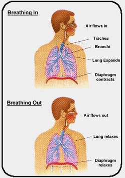
Muscles involved in Gas Exchange
-
-
-
- Breathing requires the help of a muscle known as the diaphragm.
- The diaphragm is a large, sheet-like muscle at the bottom of your chest cavity. It helps you exhale and inhale by moving up and down, respectively.
- When your diaphragm contracts (moves up), you inhale air.
- When your diaphragm relaxes (moves down), you exhale air.
- Without your diaphragm, you lungs couldn’t fill up with fresh air or push old air out.
-
-
Blood Flow
For gas exchange to occur in the lungs and the rest of the body’s tissues, blood must flow continuously through the tissues. The heart pushes blood through the tissues and provides a constant force for blood flow to occur. The heart provides enough force to propel the blood through the arteries and veins in the body. The arteries entering tissues, called arterioles, can constrict (become more narrow) or dilate (become relaxed and less narrow) to change the amount of blood flowing to an area. If an arteriole constricts, less blood is available for the tissues it supplies. If an arteriole dilates, more blood reaches the tissues it supplies. Why is it useful for the arteries to change size? Can you think of situations where certain tissues may need more or less blood flow?Blood Pressure
Blood pressure is a measure of the force needed for blood to move through the tissues.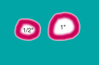
-
- This pressure depends on the amount of blood in the body, the diameter of the blood vessels, and how hard the heart is pumping blood.
- Resistance in the circulatory system is caused by the blood rubbing against the walls of the blood vessels as it flows through them. This rubbing produces a force, called resistance, opposite the blood flow.
- A large vessel is less resistant than a small blood vessel because relatively less blood rubs against the walls of the blood vessel, while a small blood vessel is more resistant because it has a smaller area for the blood to flow through. This means that more blood rubs against the walls of the vessel and it slows blood flow.
-
- In any one capillary, this resistance is an advantage because the slowed blood flow has more time for gas exchange to occur.
- When an arteriole dilates, the diameter almost doubles. When a vessel’s diameter increases, the blood flow increases by four times the original amount.
- This is like the difference between a water hose with a 1/2″ inner diameter and a 1″ inner diameter water hose. Under the same pressure, the 1″ hose will have 4 times the flow of the smaller hose.
- With decreased resistance, more blood can flow to the tissues that need more nutrients and gas exchange. For example, under conditions of moderate exercise, blood flow to skeletal muscles can increase by up to 10 times, because the arterioles in that area become dilated and supply more blood to the muscles.
- Under conditions of little resistance, the heart does not have to work as hard to move blood into the tissues.
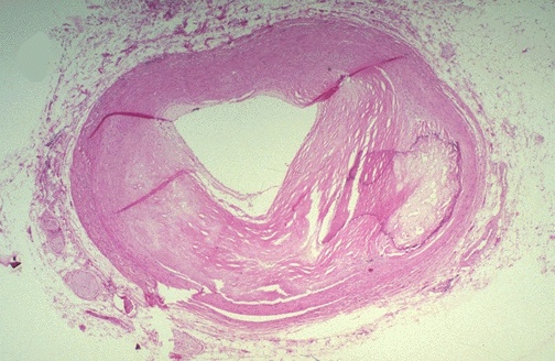
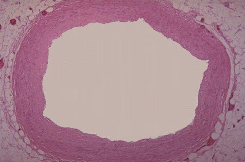
- Many people have blood pressure that is too high (called hypertension.) One known cause of hypertension is atherosclerosis. Atherosclerosis is a condition in which the walls of the blood vessels become thick and stiff, reducing their flexibility and ability to dilate.
- Eventually fats and cholesterol build up and reduce the diameter of the blood vessels, making it very difficult to for blood to flow through the vessel. In some cases, blood is unable to flow at all. This puts a tremendous amount of stress on the heart and can lead to heart failure.
- When an artery supplying the brain or heart is blocked, tissue damage can occur very quickly and if not treated immediately, is permanent and can lead to death.
A little flap covers the trachea each time you swallow to direct food and drinks to go down the esophagus, a tube that leads to the stomach. When food and drinks find their way into the trachea, you’ll know it immediately! You’ll begin coughing in order to push the food back out of the trachea to allow air to pass through uninhibited.
What happens when a person laughs so hard, that milk can come out of their nose?
-
- As the trachea approaches the lungs, it branches into two tubes. One tube leads to the right lobe of the lungs, and the other tube leads to the left lobe of the lungs. These tubes are known as the bronchial tubes. The bronchial tubes enter the lungs and begin branching into smaller and smaller tubes.
- These smaller tubes are called bronchioles. The walls of the bronchioles contain smooth muscle that is used to dilate or constrict the lungs depending upon the body’s need for oxygen. The bronchioles continue the branching process until they reach small, thin sacs called alveoli. The function of the lungs depends primarily on these tiny structures.