
A&M Researcher Receives Grant from the American Heart Association
The human body has the amazing ability to respond to disease and trauma by growing new blood vessels that in turn supply injured or diseased tissues with increased amounts of oxygen and nutrients. In some cases, such as with cancer, malignant cells can actually “hijack” this process and force the growth of new blood vessels to feed a tumor. Many therapies and treatments currently in practice attempt to destroy these new blood vessels in hopes of “starving” the invading cells.
 New research funded by the American Heart Association and conducted by Dr. Gonzalo Rivera, assistant professor in veterinary pathobiology at Texas A&M College of Veterinary Medicine & Biomedical Sciences, aims to understand the formation of new blood vessels (called angiogenesis) with the hope to discover ways to prevent their growth before it happens.
“Our laboratory focuses on cellular physiology with particular emphasis on the processes that determine cell shape, movement, and invasion,” said Rivera. “These are fundamental aspects in development and disease that are dependent on changes in the cytoskeleton, a meshwork composed of many copies of building blocks that fit together – just like the Legos blocks that children play with – to form cellular structures and appendages that aid in movement. While we address fundamental questions concerning the biology of the cytoskeleton, we also strive to instill a translational character to our research – the applicability of new knowledge to the better understanding of disease progression and potential cures. As such, our long-term goal is to understand the role of the cytoskeleton in biomedically relevant phenomena, including angiogenesis and tumor progression and metastasis.”
Research in the Rivera lab relies on a variety of tools and techniques ranging from molecular and cellular biology, to proteomics (which combines molecular genetics and biochemistry to investigate protein function at the cell level), to high-resolution optical microscopy. Rivera’s team is able to combine these techniques to better observe cell movement and growth.
“Using these approaches and with the support of this new grant from the American Heart Association, we will address the outstanding critical issues of how endothelial cells (cells lining our blood vessels) integrate multiple clues from the surrounding tissues and translate them into internal biochemical signals; and how cytoskeletal changes induced by the these biochemical signals influence vascular formation and organization,” notes Rivera.
The results from this research will expand the knowledge that researchers have about how the body responds to trauma and disease at the cellular level, providing new opportunities to develop therapies for diseases such as cancer, diabetes, and traumatic injury. Dr. Rivera credits the success in securing these funds to the hard work and dedication of team members Dr. Sankar P. Chaki (Postdoctoral Research Associate) and Ms. Rajeswhari Yog (Research Assistant), as well as the highly supportive environment of the CVM, and particularly, the Department of Pathobiology and the Image Analysis Laboratory.
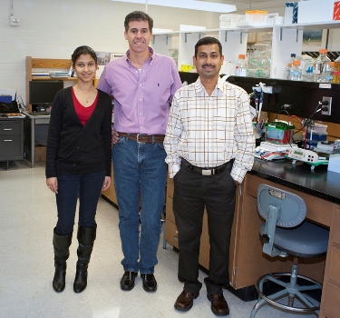
CUTLINE FOR ABOVE PHOTO: Dr. Gonzalo Rivera (center) is assisted in his lab by Dr. Sankar P. Chaki (Postdoctoral Research Associate, right) and Ms. Rajeswhari Yog (Research Assistant, left) in investigating the role of cytoskeletal proteins in the development and function of blood vessels in the hopes that the answers they find can one day be used to fight diseases like cancer and traumatic injury.

