
“Miracle Foal” Survives Traumatic Brain Injury
COLLEGE STATION, TX -After a month of surgeries and careful treatment, Reno, a three month old Quarter Horse/Welsh Pony colt, is going home. On June 19th, Reno was brought to the Texas A&M College of Veterinary Medicine & Biomedical Sciences (CVM) Veterinary Medical Teaching Hospital (VMTH) after he sustained blunt force trauma to the head, with bone chips embedded in his brain. Today, Reno is finally going home thanks to the teamwork and dedication of multiple specialists at the CVM.
 The owners, Jody Baton and her daughter Whittany of Kilgore, Texas, brought Reno to the CVM for treatment as quickly as possible after the referring veterinarian, Dr. Robert Thoni identified a skull fracture with an x-ray image. Baton is no stranger to the Large Animal Hospital, 11 of her horses have previously been brought here for specialty treatment or surgery.
“It’s like a family reunion when I come in. These veterinarians are angels,” Baton said.
When Reno arrived at the CVM, he was very depressed and had difficulty walking. Dr. Keith Chaffin, professor of equine internal medicine, was assigned to lead Reno’s case. After rapid stabilization therapy, Reno was immediately sent for MRI and CT scans.
“The magnitude of brain swelling was much worse than we predicted, and the CT scan showed more than 20 bone chips embedded in the brain,” Chaffin said. “We couldn’t have known the extent of Reno’s injuries if it weren’t for the new Diagnostic Imaging and Cancer Treatment Center. What we can now do with brain and head injuries is state-of-the-art and we can better diagnose and develop therapeutic treatments, and Reno is a great example of that.”
Reno’s only chance for survival was surgery. Baton did not hesitate in making the decision to proceed.
“He is an extraordinary colt, and we didn’t think twice about agreeing to surgery because we knew he would be in good hands,” Baton said. “Besides, how do you put a price tag on a family member?”
Dr. Joseph M. Mankin, clinical assistant professor of neurology and neurosurgery in the CVM Small Animal Hospital, performed the surgery.
“What sets the CVM apart is our access to other specialties, the team effort for Reno has been phenomenal,” Chaffin said. “This type of surgery was somewhat unchartered territory for the Large Animal Hospital, and Dr. Mankin did an excellent job.”
Mankin removed about 25 bone fragments, a piece of skin with hair, and most alarming for the team, a pus pocket. The pus pocket indicated infection was already present, and an increased possibility for more extensive infection post-operatively. But an infection in the brain wasn’t the team’s only concern after surgery, Reno reacted violently as he awoke from anesthesia. The team was forced to anesthetize him again. This scenario repeated itself twice more and on the third attempt Reno was able to wake calmly. However, Reno tore the top side of his urinary bladder during the process, and needed surgery to repair the tear. Dr. Carolyn Arnold, assistant professor of equine soft tissue surgery, led Reno’s second surgery that week.
Reno’s bladder surgery and recovery went well, and there were no violent episodes. But post-surgery, colic and a fungal infection of the tongue slowed his recovery. Gastric ulceration was the cause of colic and he responded to therapy with a proton pump inhibitor. The tongue infection was caused by candidiasis, and was successfully treated with antifungal agents.
“Reno just had crisis, after crisis, after crisis,” Chaffin said. “He was a challenging case, but a very special little foal.”
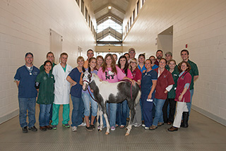
A month of careful monitoring and the teamwork of the specialists at the CVM Large and Small Animal Hospitals allowed Reno to make a full recovery. Baton, who stayed by Reno’s side almost the entire month, was joined by her two daughters and mother to take Reno home, as part of their family.
“We are just so very thankful for everything A&M has done for Reno and our family,” Baton said. “We call him our miracle foal.”

