

Cells contain several kinds of organelles. An organelle (ôrɡəˈnel) is any of a number of organized or specialized structures inside a living cell. Ribosomes are among the most crucial organelles in cells. Here, we tell the story of how ribosomes were discovered.
Organelles are important to you. For instance, from time to time, you will get sick. We usually blame sickness on some problem with our organs. Examples include lungs when we get the flu; or muscle when we have a bruise; or skin when we get sunburned. All disease begins with damage to one or more organelles within cells. The organelles allow cells to run smoothly when we are well.
Well now, you might ask, “Why should I care about how an organelle was discovered?” First, the discoveries of the various organelles tell a story about how scientists make discoveries. The story typically tells about how gaps in existing knowledge lead to new questions that motivate the scientist to develop a plan for testing.

Before we begin Dr. Palade’s story, it helps to see how centuries of work on other organelles came before Palade. It all began with Robert Hooke, who invented a microscope that he used to magnify objects enough so that cells became visible. He worked with cork, which is actually dead tissue from the cork tree. What he saw were empty circles that he named “cells” because they reminded him of a prison cell that holds prisoners. But that raised a question that Hooke could not answer: “What did these cells contain when the plant was alive?”
Over the years, many other people with better equipment and ideas found the answers. To provide a little historical perspective, here is a list of major organelles and when they were discovered.

Meet the Scientist,
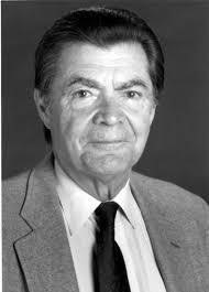
George Palade
1912-2008
Dr. George Palade is considered by many to be the father of modern cell biology. He developed a unique way to separate the various organelles in cells. His method was valuable because it allowed organelles of a cell to be separated from each other with little damage. As a result, it was possible to learn the normal function of organelles. Dr. Palade also discovered ribosomes and their function and provided us with new information about other cell organelles. Because he could purify ribosomes without damage, he was able to discover their function in protein synthesis. This is turn helped other researchers find treatments for such diseases as cancer, anemia, bone marrow failure, and various genetic diseases.

George Palade was born in 1912 in Jassy (Iasi), the old capital of Moldavia, Romania. His father was a philosophy professor, and his mother was a teacher. In 1930, he started medical school in Romania at the University of Bucharest. During medical school he discovered that he was more interested in basic science than in medicine. After medical school, he started work on a PhD degree studying dolphin kidneys. He was trying to understand how the kidneys of marine mammals, such as dolphins, are able to get rid of all of the salt that is found in sea water. Little did he know that he was about to become a pioneer in cell biology.
During World War II, he served in the medical corps of the Romanian army. After the war, he came to the United States to continue his studies. In 1952, he became a citizen of the United States. While working for a few months in the biology laboratory of Robert Chambers at New York University, Palade met Dr. Albert Claude who was giving a seminar on his work with electron microscopy (EM). This technology magnified organelles enough to be seen. That stimulated Dr. Palade’s interest in organelles. After the seminar, he chatted with Claude and somehow managed on the spot to get invited to work in Claude’s lab at The Rockefeller Institute for Medical Research.

During the 1950s, Dr. Palade discovered ribosomes and their function. He also defined the fine structure of mitochondria (Figure 1).

 Dr. Palade was interested in more than just the structure of cell organelles. He also wanted to know their function. Dr. Palade needed a source of tissue that would provide living cells that he could study. He chose the guinea pig. He also needed a tissue that was always “busy” making secretions. For that, he chose the pancreas. The pancreas is very “busy” making gland secretions. Palade spent special effort studying ribosomes, because they contained a mixture of RNA (ribonucleic acid) and the proteins that they make.
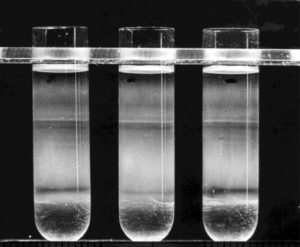
At that time, the usual method to study the parts of a cell involved homogenizing (həˈmäjəˌnīz ing). This process breaks cells open to release their contents. Spinning (centrifuging) tissue samples at high speeds separates the organelles according to their density. The heavier particles are thrown toward the bottom of the test tube, while lighter-weight particles form the upper layers (Figure 2).
This process tore the organelles apart, and the resulting fractions were not very pure. Dr. Palade wanted to isolate cell organelles without damaging them so that he could analyze their biochemistry. He and two colleagues in Claude’s lab modified the usual method of separating cell components by centrifuging homogenates in a sucrose medium. Scientists still use this method to extract specific molecules from plant or animal tissue.
The fractionation occurs in a tube prepared by layering progressively less dense sucrose solutions one upon one another without mixing. For example, the first layer of highest concentration is placed carefully in the bottom of a centrifuge tube. Then, in succession, layers of decreasing concentration are added carefully without mixing. Then cell homogenate is placed on top and centrifuged at very high speeds. Each cellular component begins to move down through the gradient and eventually reaches a position where the density of the organelle equals the density of the sucrose layer (Figure 3).

Dr. Palade continued to study the function of organelles, including the endoplasmic reticulum, the Golgi complex, the secretory granules, and the cell membrane. His love of Roman history and Latin, which is the basis of many words used in biology, were useful in helping him name many of the cell structures he identified.
Dr. Palade had a very long career and became a Dean at the University of California at San Diego School of Medicine.
Sources:
- George E, Palade. The Nobel Prize. https://www.nobelprize.org/prizes/medicine/1974/palade/auto-biography/
- Hogeboom, George H, Schneider, Walter C., and Palade, George E. (1948). Cytochemical studies of mammalian tissues: I. Isolation of Intact mitochondria from rat liver; some biochemical properties of mitochondria and submicroscopic particulate material. J. Biol. Chemistry. 172: 619-635.
- Palade G.E., et al. A small particulate component of the cytoplasm. J Biophys Biochem Cytol, 1955, 1: 59-68. http://jcb.rupress.org/cgi/reprint/1/1/59
- Schekman, Randy W. George E. Palade (1912-2008). Science. 31 Oct 2008: Vol. 322, Issue 5902, pp. 695. DOI: 10.1126/science.1167174
Author: W. R. Klemm

Do you know that different cells have certain unique proteins that have unique jobs? Muscles have proteins that contract, enabling the muscle cell to contract. Pancreas cells make the protein insulin, which regulates blood sugar levels. There are unique cells in the part of the brain called the hypothalamus that make small unique proteins that trigger the release of pituitary hormones. There are proteins in nerve cells that work to create new axon terminals and synapses that store your memories? How many other examples can you think of cells with specific proteins?