
Research at Texas A&M uncovers new facets of blood vessel formation
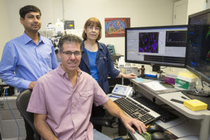
COLLEGE STATION, Texas – Researchers at Texas A&M College of Veterinary Medicine & Biomedical Sciences (CVM) are a step closer to understanding how blood vessels form. In a study published in Molecular Biology of the Cell and highlighted on the journal’s cover, Drs. Sankar P. Chaki, Rola Barhoumi, and Gonzalo Rivera identified the contribution of Nck to morphological changes involved in early steps of blood vessel formation. The findings suggest Nck, a molecule that integrates key cues from the tissue microenvironment, plays an important role in coordinating the behavior of the endothelial cells that line blood vessels.
Although Nck was known to influence the development of the vascular network, its specific role in the molecular and cellular processes involved in blood vessel formation had remained undetermined. This study showed that Nck promotes the organization of endothelial cells into a well-connected network of tubes resembling the vascular tree. Results from this investigation highlight that Nck plays a critical role in the establishment of cell-to-cell contacts and the polarized organization of endothelial cells.
“Polarity in animal cells is evidenced by the asymmetrical distribution of organelles and molecular components, and by virtue of such polarity, cells can perform specialized functions. Not surprisingly, loss of cell polarity is associated with various disease states. Polarity of endothelial cells is critical for the delivery of oxygen and nutrients to the tissues, and the removal of metabolic waste,” explained Chaki, the lead author in this publication. He added “Highlighting the molecular and cellular processes underlying vascular formation is important not only from a development standpoint–the complex body plan of an animal just would not develop without a robust, functional vascular network–but also to understand key processes in well-being, such as wound healing and tissue repair.”
As expected, the analysis of important biological problems at the cellular and molecular levels presents significant technical challenges. “We combined a three-dimensional tissue culture system that recreates key features of the tissue microenvironment with state-of-the-art optical imaging techniques that enable the capture of cellular and molecular processes with high spatial and temporal resolution”, emphasized Dr. Rola Barhoumi, co-author in the study and associate director of the Image Analysis Laboratory.
By understanding the fundamental processes involved in blood vessel formation, researchers may also be able to develop safe and effective treatments to regulate blood vessel formation in disease. For example, this is very relevant to cancer therapy. “Once solid tumors reach one to two millimeters in size, they begin secreting factors that stimulate the development of infiltrating blood vessels,” Rivera said. “The newly formed vasculature provides nutrients and factors that, in turn, promote tumor growth. Additionally, blood vessels in tumors are often dysfunctional and leaky, which can allow cancer cells to spread to other parts of the body. Therefore, effective treatments would work to repair existing vessels as well as stop blood new blood vessels from reaching the tumor.”
“The article and journal cover are a tribute to Drs. Rivera, Chaki, and Barhoumi, who have combined sophisticated molecular biology approaches and advanced imaging tools to investigate the formation of endothelial tubes that mimic blood vessel formation,” said Dr. Robert Burghardt, associate dean for research and graduate studies. “This work is very significant because of its translational potential, since these findings suggest that targeting key molecular pathways regulating the cytoskeleton is an emerging approach to therapies that require the control of blood vessel formation.”

