
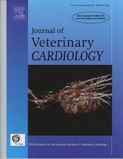
Computed Tomography Image Selected for Journal Cover
COLLEGE STATION, TX – Medical imaging saves lives. Mammograms find breast cancer, ultrasounds show the extent of injuries after an accident, and MRIs detect brain aneurysms. Diagnostic imaging is also an increasingly important tool in veterinary medicine.
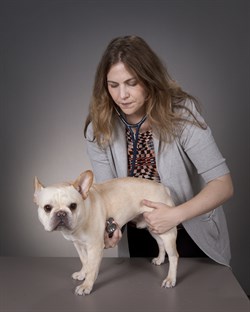
When a canine patient with a heart murmur was referred to the Texas A&M College of Veterinary Medicine & Biomedical Sciences (CVM), a team led by Dr. Ashley Saunders used a computed tomography (CT) scanner and a software program to create a three dimensional model of the dog’s heart. The images were used to help the team develop a detailed treatment plan for a complex problem: correcting an uncommon heart defect that was causing the murmur and associated health problems.
The dog’s case was highlighted in a recent issue of the Journal of Veterinary Cardiology. Due to the unique aspects of the case; successful treatment; and the overall clarity and quality of the accompanying images, one of the three dimensional reconstructed images of the heart defect was selected as the journal cover.
“The images allowed us to visualize the dog’s anatomy, identify the abnormalities, and make quick decisions about his surgical plan,” said Saunders, an associate professor in the Department of Small Animal Clinical Sciences (VSCS). “The information from these images helped us perform a successful procedure that resulted in an excellent outcome for the patient.”
“At Texas A&M, we work to put the very best tools in the experienced hands of our clinicians,” said Dr. Eleanor Green, Carl B. King Dean of Veterinary Medicine. “Our Diagnostic Imaging and Cancer Treatment Center is a leading-edge facility that enhances our ability to treat difficult cases and to provide an advanced level of care. The article and cover photo are a tribute to Dr. Saunders and to our excellent cardiology team. The collaborations they establish, and the advanced technology they use in their daily patient care, allows them to make a positive difference in the lives of those we serve.”
The CT scanner used for the angiogram study is housed within the
Diagnostic Imaging and Cancer Treatment Center at the CVM, one of the only facilities of its kind. The 3D reconstruction was performed with software at the Texas A&M Institute for Preclinical Studies (TIPS). Access to these resources and this technology aids veterinarians at the CVM in providing the very best patient care and saving animals’ lives.
“Being featured on the journal’s cover is a notable accomplishment,” Dr. Sandee Hartsfield, professor and head of VSCS noted. “Only a limited number of submitted papers are recognized with a cover image, and both the articles and the images must be of the highest quality.”

