
Texas A&M Veterinarians Create New Strategies For Treating Ear Issues In Kangaroos
Story by Megan Myers, VMBS Communications
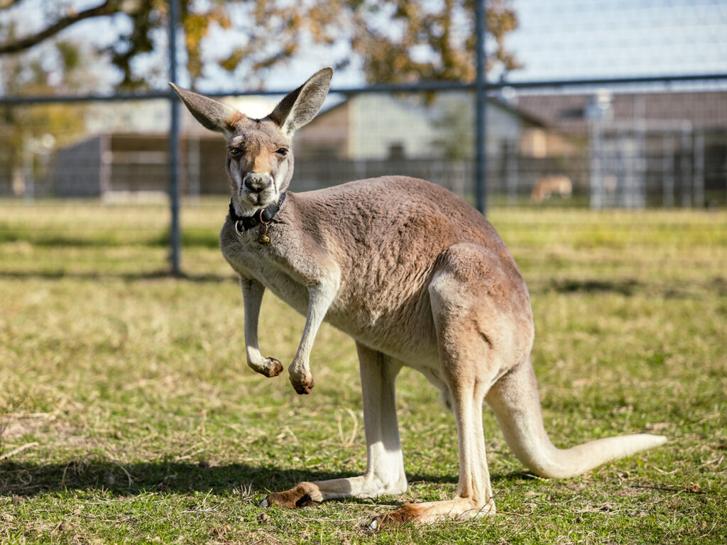
Photo by Jason Nitsch ’14, Texas A&M University Division of Marketing and Communications
Diego the red kangaroo has an important job at the Texas A&M Winnie Carter Wildlife Center. In addition to teaching veterinary students about kangaroo care and medicine, he serves as a “seeing eye” kangaroo for his blind “roo mate,” Murdock.
When Diego’s caretakers at the wildlife center noticed that he seemed to be experiencing discomfort in his right ear, they knew it was important to get him back to tip-top — or, more aptly, hip hop — shape so he could continue his normal life with his friend.
But because of the minimal information published about ear issues in kangaroos, Diego’s veterinary team at the Texas A&M School of Veterinary Medicine & Biomedical Sciences’ Small Animal Teaching Hospital (SATH) had to start from scratch in figuring out what equipment and strategies to use.
Look Before You Leap
Diego’s ear problem became apparent not long after his arrival at the wildlife center in 2022.
“You could always tell which one was Diego because his right ear was bothering him a lot; he’d hold it to the side and scratch at it and sometimes shake his head a little bit,” said Dr. Alice Blue-McLendon, director of the wildlife center and a clinical associate professor.
During Diego’s first visit to the SATH, his veterinarians in the Dermatology Service, which deals with a wide range of skin, ear and allergic conditions, could tell that something was wrong with his ear canal, but because the canal was so small, none of their equipment could fit inside.
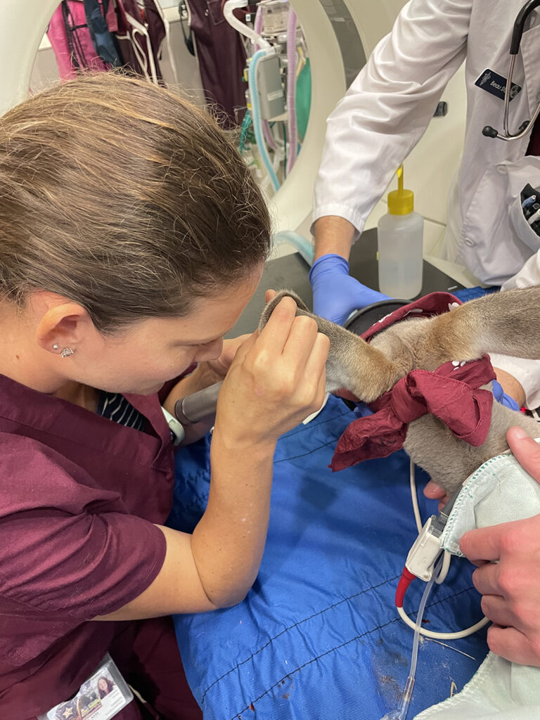
Photo by Dr. Alice Blue-McLendon
“Kangaroos have an ear canal that is relatively firm, or cartilaginous, so there’s not a lot of give to it. It also has what we call a ‘double bump,’ which is almost like two stair steps, so it’s not a straight shot to the bottom of the canal,” said Dr. Christina Gentry, a clinical assistant professor and veterinary dermatologist.
After giving the kangaroo some time to grow, Gentry was finally able to address the problem.
“We found a keratin buildup called a ceruminolith, which is almost like a rock of wax,” Gentry said. “It sometimes occurs because a tiny object, like a grain of sand or a piece of stick, gets into the ear. The body builds up more and more wax around it and then the ear canal becomes inflamed and narrows, not letting the growing wax ball exit.”
While the condition is commonly seen in cats and sometimes in dogs, there is little documentation about it occurring in kangaroos. Likewise, there is hardly any published literature about the species’ ear canal anatomy, making Diego’s health journey a challenging expedition.
Even with the challenge posed by Diego’s anatomy, Gentry and her team were determined to find a way to remove the blockage.
“Our concern was that if he ended up deaf or had chronic pain in his ears, he would not be able to behave normally and, therefore, could not help his friend,” Gentry said.
Jumping For Joy
After using a video otoscope, a small camera that magnifies images, to see the ceruminolith inside Diego’s ear, Gentry wanted to make sure there was not any other issue at play, so she ordered a CT scan to check on the kangaroo’s middle ear, or the area right behind the eardrum.
“The CT showed that his middle ears were normal and that all the disease was confined to the external ear canal,” she said. “While he was still asleep, we used the video otoscope’s guidance to remove the majority of the wax blob with sterile saline flush and tiny forceps.”
Because the canal was so inflamed, the veterinarians still couldn’t see the eardrum even once the blockage was removed, so they sent Diego home with some medications and set a recheck appointment for four weeks later, once the swelling had time to go down.
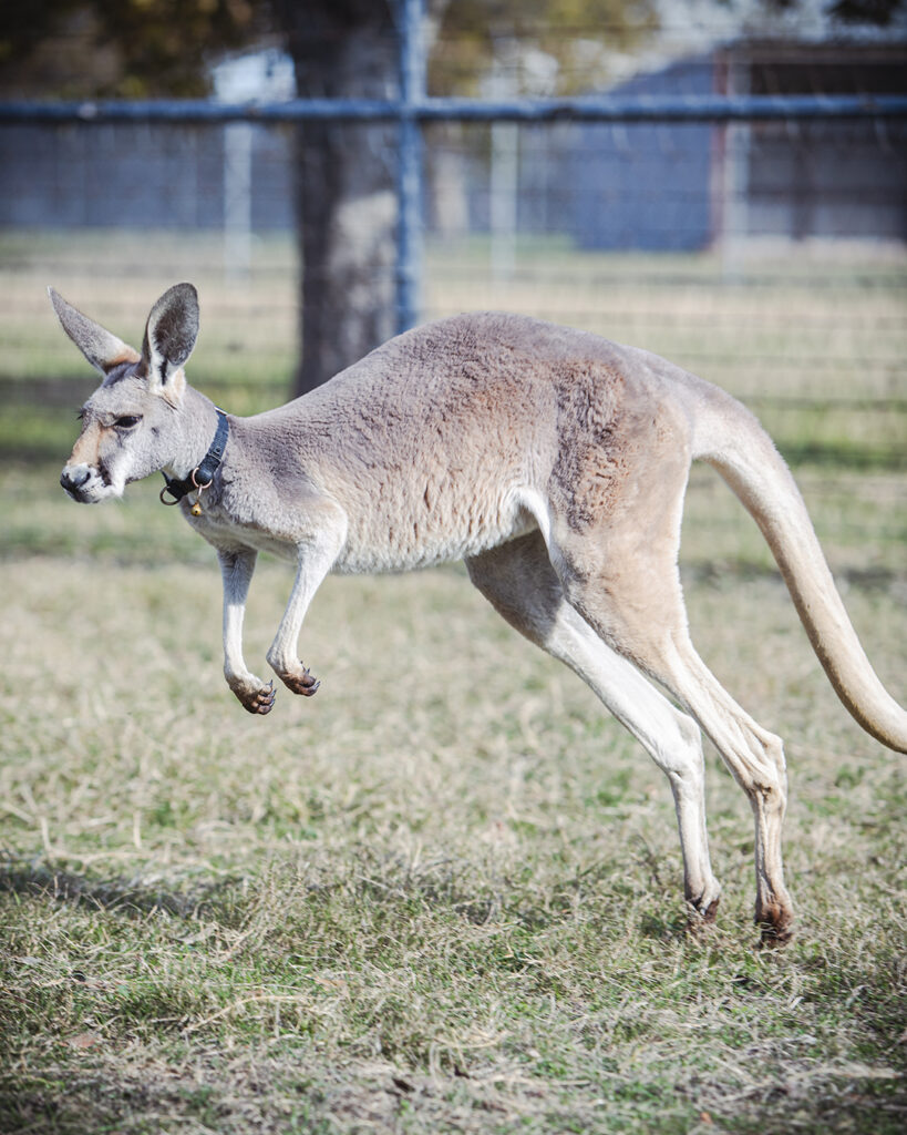
Photo by Alyssa Moore ’27, Texas A&M School of Veterinary Medicine & Biomedical Sciences
While they hoped they had solved the issue by removing the ceruminolith, there was a possibility that the blockage had caused permanent damage that could even require surgical removal of the entire ear canal.
But, to the joy of Diego’s care team, the follow-up visit found that the kangaroo was healing well.
“The canal looked much better and was not infected, and we were able to see down to the level of the eardrum,” Gentry said. “We are now moving to maintenance therapy with ear drops twice a week for the next few months.”
Healing Abounds
Since his procedure, Diego has been shaking his head less and holding his ear up more, indicating to his caregivers that he is feeling better.
Because Diego’s case was so unique, his veterinary team is hoping to share its findings and encourage future research on kangaroo ear canals.
“We now know that as kangaroos get older, traditional video otoscopy becomes more useful,” Gentry said. “At some point, once they hit maybe 2 years of age, you can use traditional instrumentation that you would use in a dog and cat, but below that age, you may need to acquire a small flexible endoscopic tool.”
Sharing Diego’s CT scans will also inform other veterinarians of how to best treat ear conditions in kangaroos.
“Just because a kangaroo is an exotic species doesn’t mean they can’t still be affected by some of the diseases that we see in pets,” Gentry said. “It’s helpful to have a better idea of the ear canal anatomy of kangaroos, knowing that it doesn’t look exactly like a dog’s or a cat’s.”
A Hoppy Family
Diego and Murdock arrived at Texas A&M as babies, weighing only about 12 pounds and still drinking kangaroo milk replacement, imported from Australia, from a bottle.
Because kangaroos are social animals and because Murdock has cataracts and detached retinas in both eyes that have rendered him blind, Diego’s main “job” at the wildlife center is to be Murdock’s companion.
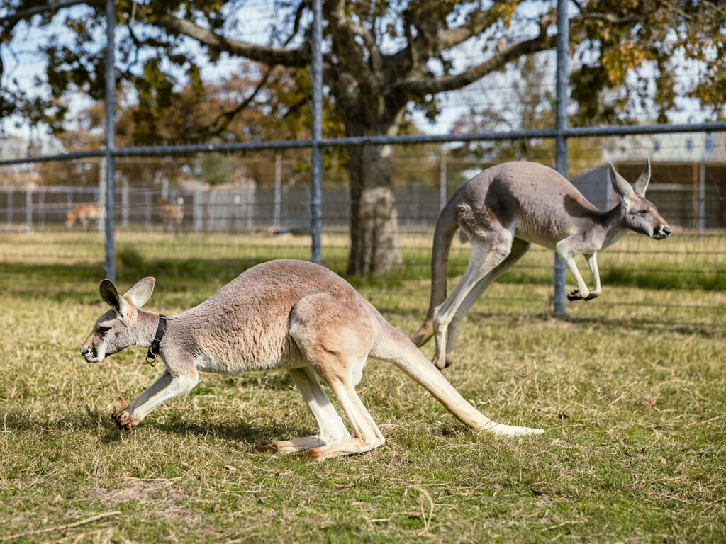
Photo by Jason Nitsch ’14, Texas A&M University Division of Marketing and Communications
“We did not want to put Murdock around our two adult kangaroos because of his blindness, so we sought a ‘seeing eye roo,’” said Blue-McLendon. “Diego wears a collar with little bells on it so Murdock can hear him and figure out where his companion is.”
In addition to serving as a sanctuary for dozens of exotic animals, the Winnie Carter Wildlife Center serves as a teaching facility for undergraduate, graduate, and veterinary students who are interested in wildlife conservation and medicine.
It’s tradition at the wildlife center to name the animals after school administrators, donors and volunteers. Diego was named after a wildlife and fisheries sciences former student who had volunteered at the center for several years and was given the honor of picking up the kangaroo and bringing him to campus.
Now that Diego’s ear is healed, he and Murdock spend their days hopping around their outdoor paddock at the wildlife center alongside their new joey friend, Suva (named after Dr. Larry Suva, head of the Department of Physiology & Pharmacology).
###
For more information about the Texas A&M School of Veterinary Medicine & Biomedical Sciences, please visit our website at vetmed.tamu.edu or join us on Facebook, Instagram, and Twitter.
Contact Information: Jennifer Gauntt, Director of VMBS Communications, Texas A&M School of Veterinary Medicine & Biomedical Sciences, jgauntt@cvm.tamu.edu, 979-862-4216

