
Texas A&M Veterinarians Leave No Stone Unturned In Diagnosing Horse’s Mysterious Colic
Story by Megan Myers, VMBS Communications
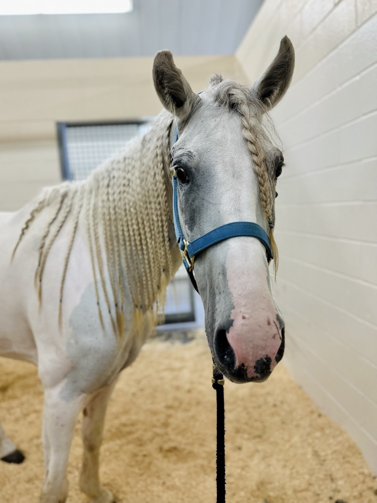
When Koche, an American Paint Horse, began experiencing recurring bouts of colic, her owner, Cedrick Harvey, was determined to discover the source of the problem because of the 7-year-old mare’s special place in his family.
“Her grandmother was an amazing Paint horse named Apache that grew up within our family,” Harvey said. “We really wanted to keep her bloodline alive, so we bred her and got Cody, who was Koche’s father. So, I’ve had Koche since her birth.”
During Koche’s first seven years, she was remarkably healthy, which made it all the more concerning when Harvey saw her begin showing signs of pain in March 2023.
When Harvey’s local veterinarian was unable to determine the cause through a physical exam, he referred Koche to the Texas A&M School of Veterinary Medicine & Biomedical Sciences’ (VMBS) Large Animal Teaching Hospital (LATH), where advanced equipment was available to help diagnose her condition.
After the two-hour drive from Crosby to College Station, Harvey put his beloved mare in the hands of the LATH’s equine veterinarians.
“When I drove up and saw the team of doctors and students standing outside waiting on her, it blew me away,” he said. “I was so pleased when I saw how much care and attention they showed her. I knew she was in great hands.”
Diagnosing The Problem
Colic is a general term for abdominal pain in horses and can have a variety of causes. No matter the specific cause, however, horses experiencing colic tend to react the same.
“They do what I call the ‘no-pants dance,’” said Dr. Dustin Major, a VMBS clinical assistant professor of large animal surgery. “They circle around and posture like they’re about to lay down, but then they never do. Koche was doing that little dance and kicking at her belly, so we knew she was in pain.”
After providing some medication to reduce her pain, Major began what is commonly known as “the colic workup.” He started by conducting a general physical exam and using a nasogastric (nose to stomach) tube to check for gastric reflux, followed by a rectal exam to palpate her internal organs.
“She didn’t have any reflux, and all I could feel on her rectal palpation was some feed in her large intestine,” Major said. “Nothing was out of the ordinary, and we found nothing that should have been making her show the signs of pain that she was exhibiting.”
Next, he performed an abdominocentesis, which involves collecting a sample of the abdominal fluid that can be tested for protein content, lactic acid concentration, and more.
But, once again, Koche’s test results revealed no answers.
After also finding no conclusive results from an abdominal X-ray, Major decided to move on to his final and most intensive option — colic surgery.
“If the horse is still in pain despite medical therapy and the cause can’t be determined, it goes to surgery because we need to get in there and find out what’s going on,” he said.
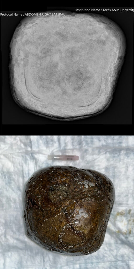
During this exploratory procedure, Major took his time studying all the abdominal organs, looking for irregularities or damaged tissue. Finally, the cause of Koche’s pain became enormously clear; there was a cantaloupe-sized stone lodged inside her large colon.
An Easy Fix
These stones, also called enteroliths, are formed in a horse’s right dorsal colon, a section of the colon that is designed to contract and pack things together.
“It’s sort of like making a pearl; the horse ingests a piece of wood, gravel, or something else and then the colon compacts minerals around it, layer upon layer upon layer,” Major said. “The stone then goes down to the transverse colon, where it lodges and backs everything up. The stone can come back out of the transverse colon and then lodge in it again, which is why these horses can be intermittently painful.”
Fortunately for Koche, the nearly five-pound stone was easily removed during the same surgery. Just as important, though, was ensuring this situation would not happen again.
After talking with Harvey, Major came to believe that the cause of Koche’s enterolith was a diet high in alfalfa, combined with the high calcium content of Crosby’s water.
While enteroliths are common in places like California, most horse owners in Texas are less familiar with the condition because of its relative rarity in the state.
“Alfalfa has a lot of calcium, so if you feed more than 50% of the diet as alfalfa, these stones are possible,” Major said. “Fortunately, it’s a preventable problem. Knowing to feed less of your horse’s diet as alfalfa is important as well as using water softeners, if possible.”
Major took an X-ray of the enterolith to try to determine what was at its center, but the results were inconclusive. It could have been anything from a piece of rope in Koche’s stall to a small rock in the pasture.
“You really want to make sure you’re watching what they’re eating and taking their diet seriously, especially if you’re as close to your animals as I am to mine,” Harvey said. “You also want to make sure you’re aware of what’s out there in the field that they could eat.”
Minutes Count
Koche’s happy ending was only possible because of her local veterinarian, the LATH veterinary team, and Harvey’s persistence in finding the source of her pain. For horses that do not receive prompt treatment, many sources of colic, including enteroliths, can eventually prove fatal.
“If you have a horse with colic and it’s not getting better, it’s better to go ahead and be aggressive on the front end,” Major said. “You’ll spend less money and have a better chance of having a successful outcome. Especially from a surgical perspective, the earlier we get into the abdomen, the better. Literally, minutes count.”
After staying at the LATH for a week, Koche was finally ready to return home.
“I really appreciated the entire team, especially a student named Valerie Ruiz (a fourth-year veterinary student at St. George’s University who completed clinical rotations at the LATH), who did a great job giving me updates on Koche,” Harvey said. “On the day I came and picked her up, all of her doctors walked out to tell me how well she was doing. It was just a top-notch experience.”
###
Note: This story originally appeared in the Fall 2023 issue of VMBS Today.
For more information about the Texas A&M School of Veterinary Medicine & Biomedical Sciences, please visit our website at vetmed.tamu.edu or join us on Facebook, Instagram, and Twitter.
Contact Information: Jennifer Gauntt, Director of VMBS Communications, Texas A&M School of Veterinary Medicine & Biomedical Sciences; jgauntt@cvm.tamu.edu; 979-862-4216

