Texas A&M Veterinary Specialists Collaborate On Rare Heart Procedure To Save Young Tabby Cat
Story by Megan Myers, VMBS Communications
At only 5 months old, Whiskey the kitten was diagnosed with congestive heart failure, a condition with potentially deadly consequences.
But thanks to committed owners and a talented veterinary team willing to try a procedure in a way never done at the Texas A&M School of Veterinary Medicine & Biomedical Sciences’ (VMBS) Small Animal Teaching Hospital (SATH), Whiskey got a second chance at life.
Discovering The Problem

Whiskey entered Vicki and Chris Hartnett’s lives in June 2020 on one of their daily walks near a heavily wooded area in their hometown of Spring.
“We were just walking past an empty lot and I’m chattering away, and, all of a sudden, Chris stopped and said he heard a little meow,” Vicki said.
The couple spotted a tiny orange tabby, about 5 weeks old, running out of the forest, straight toward them; although they looked for a long time, they were never able to find its mother, or any other cats, nearby.
Taking the kitten home with them, they quickly fell in love with Whiskey, named for his golden color, and decided to keep him.
But trouble struck in September when Whiskey began acting sick and vomiting repeatedly. Their local veterinarian discovered a heart murmur and suggested the couple take Whiskey to the SATH’s Cardiology Service.
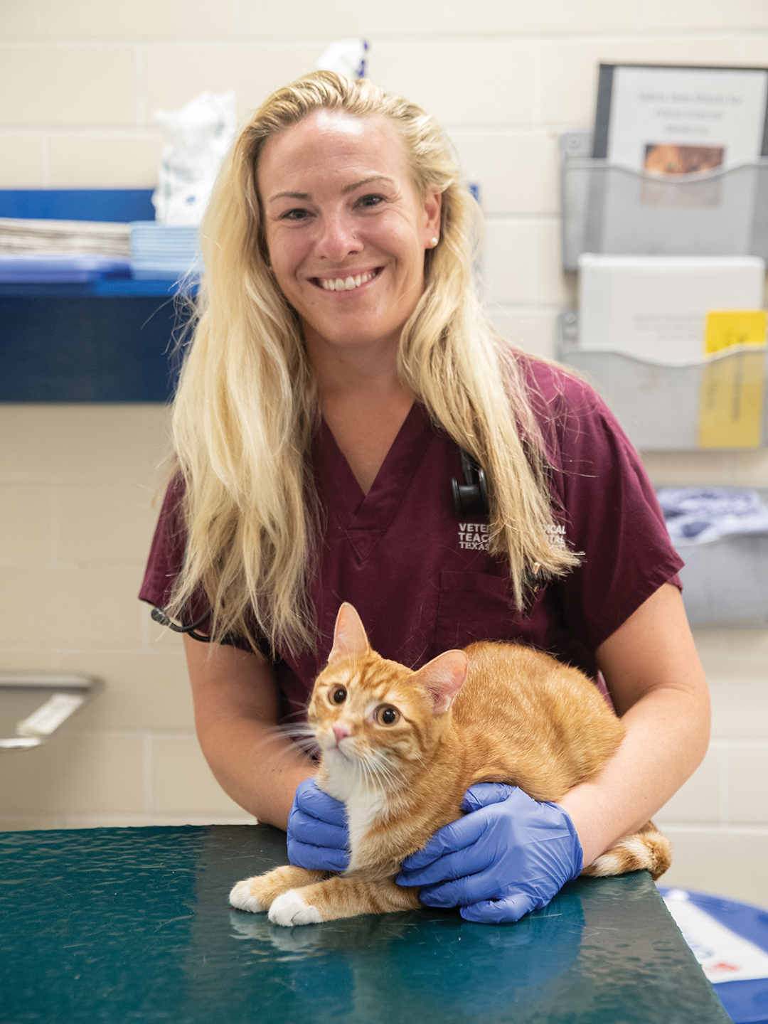
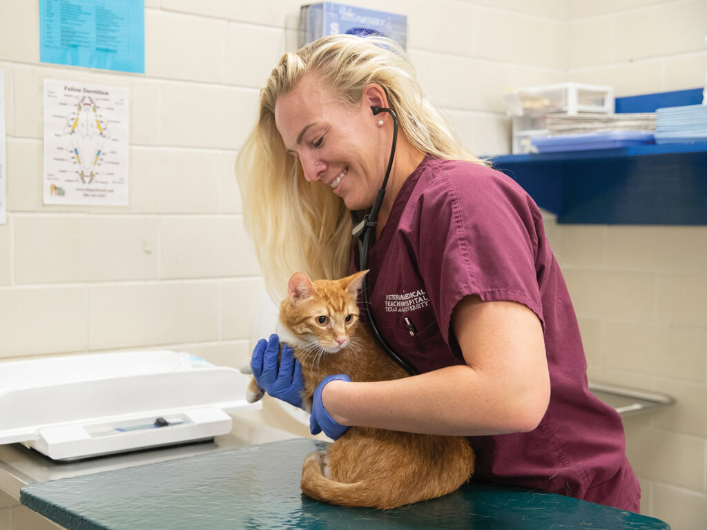
“Whiskey’s heart murmur was so loud that his actual chest wall vibrated with it,” said Dr. Caitlin Stoner, a former SATH cardiology resident. “The louder the heart murmur is, the more likely it is to be pathologic (diseased) or something else significant, and Whiskey’s murmur was truthfully about as loud as it could get.”
After a series of tests with ultrasound and chest X-rays to look at Whiskey’s heart, Stoner and VMBS professor and cardiologist Dr. Ashley Saunders found that what was causing the heart murmur was much worse than expected.
“Whiskey had a ventricular septal defect, which is a hole in the wall between the two chambers of the heart that allowed blood to flow from one side to the other; it shouldn’t normally do this,” Stoner said. “We see this fairly commonly in cats, but the thing about Whiskey’s that was so significant was the size of it — it took up a huge chunk of his heart wall.”
This hole wasn’t the only problem with Whiskey’s heart — he also had mitral valve dysplasia, meaning the valve on the left side of his heart was formed abnormally, could not open or close correctly, and was leaking significantly.
“Those two problems together led him to have severe heart enlargement at a very young age and early onset congestive heart failure,” Stoner said.
While all of the blood in Whiskey’s heart should have been circulating throughout his entire body, some of it was instead only going from the right side of the heart to the lungs and then to the left side of the heart and straight through the hole to the right side again in the wrong direction.
This separate blood flow that was only going between the lungs and heart, combined with the malfunctioning valve, was filling the left half of Whiskey’s heart with extra blood and causing it to swell. The abnormal blood flow through the hole was also causing the loud murmur.
“When those chambers of the heart max out on capacity for what they can hold, blood can back up to the level of the lungs and can actually put fluid within the lungs, causing breathing difficulties,” Stoner said.
Despite looking at a very grim situation, Whiskey’s owners refused to give up hope.
When the SATH team suggested a rare procedure that could be performed in a manner they had never done before, but that had the potential to greatly improve Whiskey’s lifespan and quality of life, the Hartnetts decided to go for it.
“From the way that Dr. Stoner explained it and all of the different hands that were going to be involved, we knew that if Whiskey was going to have a chance of success, we were in the right place,” Chris said.
Trying Something New

Before Whiskey came in for his surgery, a procedure called pulmonary artery banding, his SATH team spent a couple months planning, practicing, and reaching out to experts.
“It took us a bit of time to make sure we were as prepared as we possibly could be and had a really good plan of action,” Stoner said. “The biggest thing to emphasize about Whiskey is what a huge team effort he was; so many people came together to make that pulmonary artery banding possible.”
Finally, in mid-November, veterinarians and technicians from the Cardiology, Anesthesiology, and Soft Tissue Surgery services gathered in the operating room to try to save Whiskey’s life.
For pulmonary artery banding, a band made of a strong, synthetic resin is wrapped around the pulmonary artery and tightened to control blood flow.
“That tight belt around the artery makes it harder for blood flow to leave the right side of the heart and go out to the lungs. If it’s harder for flow to leave the right side of the heart, then less flow enters and builds up in the left side of the heart and less blood goes through the hole,” Stoner said.
Even though the hole in the heart was still there after inserting the band, the reduced fluid buildup in Whiskey’s heart meant that the hole was no longer causing the backed-up blood and resulting heart enlargement that were threatening Whiskey’s life.
As Dr. Kelley Thieman, a soft tissue surgeon and VMBS associate professor, was attaching and tightening the band, the cardiologists used a catheter inside the artery to measure the blood pressure and make sure the band was at the perfect tightness.
Once the band was tightened and the catheter removed, the surgeons used a special suture donated by St. Joseph Hospital in College Station to seal up the artery.
“We needed a small suture on a tiny needle that wouldn’t cut the vessel, but since we don’t put sutures in those great vessels all that often, we don’t carry the needle on that size of suture,” Thieman said. “St. Joseph was happy to donate it, which was fantastic.”
A Happy Ending

Once the procedure was finished and Whiskey had some time to recover, his veterinary team was overjoyed that follow-up scans showed that Whiskey’s heart had shrunk and he no longer had any fluid backing up into his lungs.
“He still has the murmur and it is still impressively loud, but under the surface, things look better,” Stoner said. “He would’ve had big problems with his heart disease in the very near future without the procedure. We reset the clock for him and were able to buy him some really good years with his family at home.”
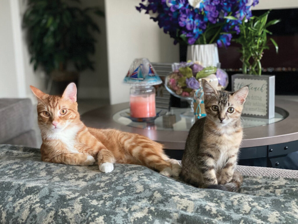
In the two years since his surgery, Whiskey has continued to do well and is living a rambunctious life like any other young cat. His owners have even adopted a little kitten “sister” for him to play with, and he has no trouble keeping up with her.
“He’s doing absolutely wonderfully,” Vicki said. “I’m just so thankful for Dr. Stoner, the entire team, and everything they’ve done for him.”
Every six months when Whiskey comes back to the SATH for a recheck, his veterinary team is reminded of how his procedure was only possible because of the teamwork and multi-service care the hospital is known for.
“The thing that I’m so proud about from Whiskey’s case is the fact that the whole hospital came together for one little cat; it was just this phenomenal team effort,” Stoner said. “It’s so neat to see how well he’s doing now and how many people are in his corner.
###
Note: This story originally appeared in the Winter 2023 edition of CVMBS Today.
For more information about the Texas A&M School of Veterinary Medicine & Biomedical Sciences, please visit our website at vetmed.tamu.edu or join us on Facebook, Instagram, and Twitter.
Contact Information: Jennifer Gauntt, Director of VMBS Communications, Texas A&M School of Veterinary Medicine & Biomedical Sciences; jgauntt@cvm.tamu.edu; 979-862-4216


