• Home
  • Clinic Login
  • Fundraising
    • Promoting Gastrointestinal Health in Companion Animals
    • Goals
    • Rob & Roxann Bilger Chair in Feline Hepatology
    • Rudy Hartrick Initiative
    • Naming Opportunities
    • Our Supporters
    • Make a Donation
    • List of Donors
  • Research
  • Service
    • Assays
    • Histopathology
    • Clinic Login
    • Frequently Asked Questions (FAQ)
    • Liability Limitation
    • Payments
    • Quality Control
    • Sample Requirements
  • More
    • Contact Us
    • Newsletters
    • Map
    • Videos
  • Staff
Skip to main content
  • Home
  • Clinic Login
  • Fundraising
    • Promoting Gastrointestinal Health in Companion Animals
    • Goals
    • Rob & Roxann Bilger Chair in Feline Hepatology
    • Rudy Hartrick Initiative
    • Naming Opportunities
    • Our Supporters
    • Make a Donation
    • List of Donors
  • Research
  • Service
    • Assays
    • Histopathology
    • Clinic Login
    • Frequently Asked Questions (FAQ)
    • Liability Limitation
    • Payments
    • Quality Control
    • Sample Requirements
  • More
    • Contact Us
    • Newsletters
    • Map
    • Videos
  • Staff
What can we help you find?
Gastrointestinal Laboratory
  • Home
  • Clinic Login
  • Fundraising
    • Promoting Gastrointestinal Health in Companion Animals
    • Goals
    • Rob & Roxann Bilger Chair in Feline Hepatology
    • Rudy Hartrick Initiative
    • Naming Opportunities
    • Our Supporters
    • Make a Donation
    • List of Donors
  • Research
  • Service
    • Assays
    • Histopathology
    • Clinic Login
    • Frequently Asked Questions (FAQ)
    • Liability Limitation
    • Payments
    • Quality Control
    • Sample Requirements
  • More
    • Contact Us
    • Newsletters
    • Map
    • Videos
  • Staff
Gastrointestinal Laboratory
  • Home
  • Service
  • Assays
  • Canine and Feline Microbiota Dysbiosis Index

Canine and Feline Microbiota Dysbiosis Index

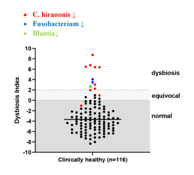
The dysbiosis index (DI) is a quantitative PCR-based assay that can be used to assess the feline (Sung et al, JFMS 2022) or canine (AlShawaqfeh M et al, FEMS 2017) fecal microbiome in individual patients. It is currently the only analytically validated assay to assess the fecal microbiome and has been used in various published clinical studies. The DI quantifies the fecal abundance of seven bacterial taxa as well as the total bacterial abundance. These bacterial taxa are commonly altered in chronic enteropathies (CE) and after broad-spectrum antibiotic use. The DI provides reference intervals for these bacterial groups and additionally calculates a single number that expresses the extent of intestinal dysbiosis (Table 1). The DI correlates negatively with species richness, i.e., a higher DI indicates lower microbial diversity.

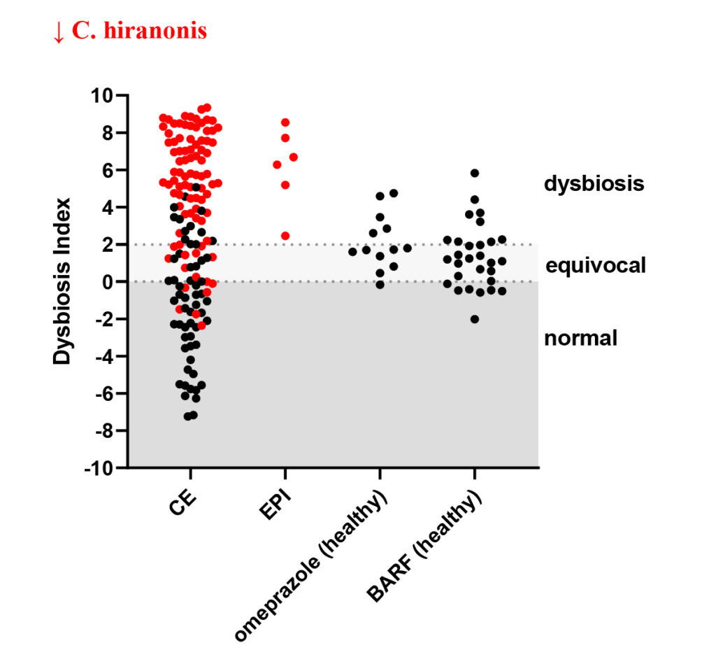
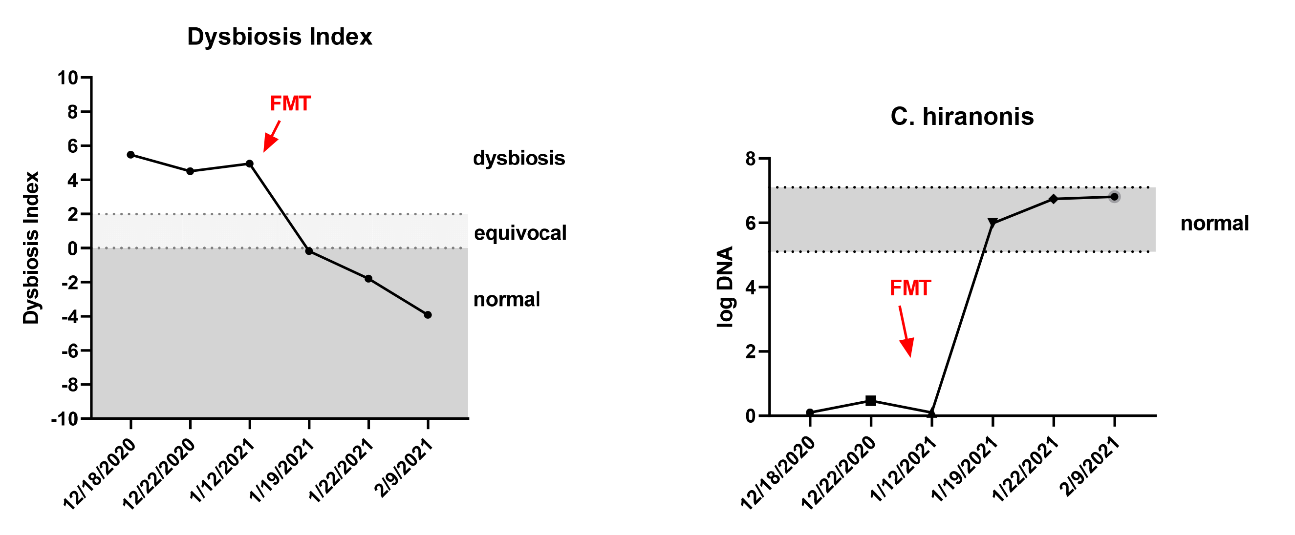
The DI also predicts, by reporting the abundance of the bile acid converting bacterium Peptacetobacter (Clostridium) hiranonis, the ability of the intestinal microbiota to convert primary to secondary bile acids (BA). Normal amounts of secondary bile acids are antimicrobial and suppress potential enteropathogens, such as C. difficile, C. perfringens, and E. coli. Therefore, a decreased abundance of P. hiranonis and decreased conversion of primary to secondary BA strongly correlates with fecal dysbiosis. An increased DI together with decreased abundance of P. hiranonis is commonly seen in EPI and CE or after antibiotic administration.

Table 1. Reference intervals for dogs and cats
Function normal in Dogs normal in Cats Change in dysbiosis
Faecalibacterium anti-inflammatory, production of SCFA 3.4 – 8.0 3.8 – 8.4 ↓
Turicibacter production of SCFA 4.6 – 8.1 4.4 – 9.0 ↓
Blautia production of SCFA 9.5 – 11.0 not measured ↓
Fusobacterium production of SCFA 7.0 – 10.3 not measured ↓
Bifidobacterium production of SCFA not measured 3.2 – 8.7 ↓
Bacteroides production of SCFA not measured 4.0 – 7.5 ↓
Peptacetobacter (Clostridium) hiranonis conversion of primary to secondary bile acids 5.1 – 7.1 4.5 – 7.1 ↓
Streptococcus overgrowth associated with dysbiosis 1.9 – 8.0 1.6 – 5.2 ↑
E. coli pro-inflammatory 0.9 – 8.0 1.4 – 7.0 ↑
Dysbiosis Index (DI) < 0
Normal DI indicating that no shifts in the overall diversity of the intestinal microbiota have been detected. If individual bacterial groups are outside the reference interval, this is suggestive of mild dysbiosis.
< 0
Normal DI indicating that no shifts in the overall diversity of the intestinal microbiota have been detected. If individual bacterial groups are outside the reference interval, this is suggestive of mild dysbiosis.
0 – 2
(DI) is mildly increased, suggesting a mild to moderate shift in the overall diversity of the intestinal microbiota
0 – 1
(DI) is mildly increased, suggesting a mild to moderate shift in the overall diversity of the intestinal microbiota
> 2
DI is significantly increased, consistent with a shift in the overall diversity of the intestinal microbiota
> 1
DI is significantly increased, consistent with a shift in the overall diversity of the intestinal microbiota
Data expressed log DNA/gram of feces
SCFA = short-chain fatty acids

Sample requirements

Approximately 1 gram of feces (approximately the size of one grape) is needed. Samples must remain cold until they reach the lab. Ship samples by overnight courier with frozen gel ice packs.  Samples can be stored in the refrigerator over the weekend if you cannot ship them by Thursday (lab personnel are not here at the weekend to receive samples). Results will typically be reported within 2 days.

See: Collections and Shipping information for fecal PCR, IFA and Bacterial Toxin Assays for more information


Increased Dysbiosis Index

The DI should be interpreted together with the abundance of the individual bacterial taxa, especially that of Peptacetobacter (Clostridium) hiranonis, as a decrease in the abundance of this species is a major contributor to an abnormal intestinal microbiome. A DI above 2 (dogs) or 1 (cats) indicates a major shift and dysbiosis with high specificity, while a DI between 0 and 2 (dogs) and 0 and 1 (cats) indicates a moderate shift in the fecal microbiome (Figure 1 and 2). Some animals with CE have a DI <0, but with some bacterial taxa outside the reference intervals, and this suggests a minor form of dysbiosis.

Before assessing the microbiome, patients should be off treatment with omeprazole and/or antibiotics. Omeprazole leads to a transient increase in the DI, but with a normal abundance of P. hiranonis. The DI normalizes within 1-2 weeks after discontinuation of therapy. Broad-spectrum antibiotics (e.g., metronidazole, tylosin) induce severe fecal dysbiosis. The microbiota typically normalizes within 2-4 weeks after discontinuation of administration in most dogs, but some dogs may have a persistent dysbiosis with lack of P. hiranonis for several months. Also, some animals on home-made diets (eg, BARF) especially those with high protein and fat but low fiber content may have an increase in the DI, but with a normal abundance of P. hiranonis.

The DI can be especially useful to evaluate healthy dogs and cats as potential donor for fecal microbiota transplantation (FMT), as a small subset of clinically healthy animals may have a shift in the microbiota. Also, animals with non-specific clinical signs (ie, lack of diarrhea and/or vomiting) may have an increased DI suggesting the presence of chronic enteropathy (Figure 4).

Therapeutic approach to microbiota dysbiosis

History and clinical presentation are important for a proper therapeutic approach to dysbiosis. Medications such as antimicrobials and proton-pump inhibitors can explain an increased DI in dogs that have otherwise no signs of intestinal disease. In most dogs the microbiota will normalize within weeks after end of therapy.

Because dysbiosis is often a component of intestinal disease, a multi-modal therapy approach addressing the underlying GI disease along with the dysbiosis should be attempted. Underlying factors predisposing dogs to dysbiosis should be corrected if they can be identified. For example, in most dogs with EPI, the microbiota  returns to normal within a few weeks or months after initiation of enzyme replacement therapy. In dogs and cats with CE, there are no clear markers that predict which treatment is best for an individual patient, and often a stepwise treatment trial is employed.

Therapeutic options for dogs and cats with intestinal dysbiosis include dietary modulation, antimicrobials, prebiotics, probiotics, synbiotics, and fecal microbiota transplantation (FMT). Each of these addresses different mechanisms and often treatments are combined for an optimal response.

Dietary modifications should always be the first treatment option. A highly digestible diet reduces undigested nutrients in the GI lumen, reducing the potential for excessive bacterial proliferation. In dogs and cats with CE, a novel or hydrolyzed protein diet reduces pro-inflammatory host responses that occur when the immune system is sensitized against the food antigen. In those patients in which a dietary change alone leads to amelioration of clinical signs the patient is diagnosed with food-responsive enteropathy (FRE). Mechanistically, the dietary change leads to gradual improvement of intestinal inflammation and dysbiosis over several months.


Fecal microbiota transplantation (FMT)

This procedure can aid in restoration of the normal microbiota. FMT is the transfer of stool from a healthy donor into the gut of a recipient via oral capsules, endoscopy, or enema. In humans, FMT has been reported to have a high success rate in the treatment of C. difficile infections. In animals, FMT is an emerging therapy, and the success appears to depend on the underlying disease process. In dogs with CE, dysbiosis is often secondary to the intestinal inflammation and structural damage. Recurrence of dysbiosis and clinical signs will occur when the underlying pathology is still present.  Therefore, in these patients, dietary and anti-inflammatory treatment of the underlying disease process is required (see above), and FMT can be considered as adjunct treatment for patients with suboptimal response to standard therapy as reported recently.

Figure 1. The Dysbiosis Index (DI) in clinically healthy dogs.
Figure 1. The Dysbiosis Index (DI) in clinically healthy dogs. A small subset of clinically healthy dogs has an increased DI and most of these dogs have a decreased abundance of P. hiranonis (in red). Some of these dogs may have persistent dysbiosis due to prior antibiotic exposure, as broad-spectrum antimicrobials (e.g., metronidazole, tylosin) have been shown to decrease P. hiranonis for weeks to months.


Figure 2: The feline dysbiosis index (DI) for healthy control (HC) cats and cats with chronic enteropathy (CE). Fifty-two of 68 (76%) cats with CE had a DI >0


Figure 2. The DI in dogs with chronic enteropathy (CE), exocrine pancreatic insufficiency (EPI), and healthy dogs on omeprazole and fed bones and raw food (BARF) diet.
Figure 3. The DI in dogs with chronic enteropathy (CE), exocrine pancreatic insufficiency (EPI), and healthy dogs on omeprazole and fed bones and raw food (BARF) diet. Some dogs with gastrointestinal disease have an increased DI which is highly associated with decreased abundance of P. hiranonis (red dots). In contrast, healthy dogs on omeprazole and BARF diets have a normal abundance of P. hiranonis.


Figure 4. Dysbiosis index in cats with chronic enteropathy separated by main clinical signs. Cats were classified based on the presence or absence of vomiting and diarrhea, regardless of other clinical signs. Cats showed only hyporexia, weight loss and/or lethargy in the group in the fourth column

Suggested FMT protocol [3]

  • Selection of donor dog – the donor should be healthy, with no history of gastrointestinal disease and recent antibiotic exposure, and should have no signs of systemic disease. The donor feces should be screened for parasites and enteropathogens. Because some clinically healthy dogs lack  P. hiranonis, which is necessary for proper BA conversion, the feces should also be evaluated using the DI
  • Storage of donor stool – stool can be fresh or stored at 4 C for up to one week in plastic bags. When feces needs to be frozen for longer storage, mixing the stool with glycerol before freezing may better preserve bacteria. Protocol: 10 grams of stool, 35 mls of saline and 5 mls of glycerol, freeze in 50 ml aliquots.
  • FMT as rectal enema – donor feces (approx. 5 grams per kg of recipient BW) is blended in 60 ml of 0.9% NaCl until liquefied. The stool is then transferred as rectal enema via a 60 ml catheter tip syringe, with an attached 12 or 14 inch French red rubber catheter. The recipient dog does not need to be sedated in most cases. If possible, do not feed and restrict the recipient dog’s activity for 4-6 hours after the transplant to lessen the chances of a premature bowel movement.


Figure 5. A dog with persistent dysbiosis after long-term antibiotic administration. FMT as enema was performed, leading to normalization of the microbiota.


References

Toresson L, Spillmann T, Pilla R, Ludvigsson U, Hellgren J, Olmedal G, and Suchodolski JS. 2023. “Clinical Effects of Faecal Microbiota Transplantation as Adjunctive Therapy in Dogs with Chronic Enteropathies—A Retrospective Case Series of 41 Dogs” Veterinary Sciences 10, no. 4: 271. https://doi.org/10.3390/vetsci10040271

Sung CH, Marsilio S, Chow B, et al. Dysbiosis index to evaluate the fecal microbiota in healthy cats and cats with chronic enteropathies. J Feline Med Surg 2022:1098612X221077876.

AlShawaqfeh MK, Wajid B, Y. Minamoto, M. Markel, J.A. Lidbury, J.M. Steiner, E. Serpedin, and J.S. Suchodolski, A dysbiosis index to assess microbial changes in fecal samples of dogs with chronic inflammatory enteropathy. FEMS Microbiol Ecol (2017) 1;93(11).

Schmid SM, Suchodolski JS, Price JM, et al. Omeprazole Minimally Alters the Fecal Microbial Community in Six Cats: A Pilot Study. Front Vet Sci 2018;5:79.

J. Chaitman, A.L. Ziese, R. Pilla, Y. Minamoto, A.B. Blake, B.C. Guard, A. Isaiah, J.A. Lidbury, J.M. Steiner, S. Unterer, and J.S. Suchodolski, Fecal microbial and metabolic profiles in dogs with acute diarrhea receiving either fecal microbiota transplantation or oral metronidazole. Front Vet Sci 7 (2020) 16;7:192.

R. Pilla, F.P. Gaschen, J.W. Barr, E. Olson, J.B. Honneffer, B.C. Guard, A.B. Blake, D. Villanueva, M.R. Khattab, M. Alshawaqfeh, J.A. Lidbury, J.M. Steiner, and J.S. Suchodolski, Effects of metronidazole on the fecal microbiome and metabolome in healthy dogs. J Vet Intern Med in press (2020).

P.R. Giaretta, R.R. Rech, B.C. Guard, A.B. Blake, A.K. Blick, J.M. Steiner, J.A. Lidbury, A.K. Cook, M. Hanifeh, T. Spillmann, S. Kilpinen, P. Syrja, and J.S. Suchodolski, Comparison of intestinal expression of the apical sodium-dependent bile acid transporter between dogs with and without chronic inflammatory enteropathy. J Vet Intern Med 32 (2018) 1918-1926.

J.F. Garcia-Mazcorro, J.S. Suchodolski, K.R. Jones, S.C. Clark-Price, S.E. Dowd, Y. Minamoto, M. Markel, J.M. Steiner, and O. Dossin, Effect of the proton pump inhibitor omeprazole on the gastrointestinal bacterial microbiota of healthy dogs. FEMS Microbiol Ecol 80 (2012) 624-36.

M. Schmidt, S. Unterer, J.S. Suchodolski, J.B. Honneffer, B.C. Guard, J.A. Lidbury, J.M. Steiner, J. Fritz, and P. Kolle, The fecal microbiome and metabolome differs between dogs fed Bones and Raw Food (BARF) diets and dogs fed commercial diets. PloS One (2018) 15;13(8):e0201279.

Chaitman J, Gaschen F. Fecal Microbiota Transplantation in Dogs. Vet Clin North Am Small Anim Pract 2021;51:219-233.

G.Q. Pereira, L.A. Gomes, I.S. Santos, A.F. Alfieri, J.S. Weese, and M.C. Costa, Fecal microbiota transplantation in puppies with canine parvovirus infection. J Vet Intern Med 32 (2018) 707-711.

S. Wang, R. Martins, M.C. Sullivan, E.S. Friedman, A.M. Misic, A. El-Fahmawi, E.C.P. De Martinis, K. O’Brien, Y. Chen, C. Bradley, G. Zhang, A.S.F. Berry, C.A. Hunter, R.N. Baldassano, M.P. Rondeau, and D.P. Beiting, Diet-induced remission in chronic enteropathy is associated with altered microbial community structure and synthesis of secondary bile acids. Microbiome 7 (2019) 31;7:126.

  • Admissions & Academics

    • DVM Professional Program
    • Graduate Programs
    • Internships & Residencies
    • BIMS Undergraduate Program
  • Departments

    • Large Animal Clinical Sciences (VLCS)
    • Small Animal Clinical Sciences (VSCS)
    • Veterinary Integrative Biosciences (VIBS)
    • Veterinary Pathobiology (VTPB)
    • Veterinary Physiology & Pharmacology (VTPP)
  • About

    • About Us
    • Awards & Honors
    • Dean's Office
  • Global Scope

    • Outreach & Service
    • Global One Health
    • International Programs
    • Education Abroad
    • VERO (VMBS + West Texas A&M Partnership)
    • Veterinary Emergency Team (VET)
  • Resources

    • Contact Information
    • Continuing Education (CE)
    • Directory
    • Facilities
    • Finance & Business Services
    • Human Resources
    • Marketing & Communications
    • Technology Services (IT)
    • UHS Counseling for Veterinary Students
    • VMBS News
  • Donors | Alumni | Clients

    • Giving
    • Alumni Relations
    • Stevenson Companion Animal Life-Care Center
    • Veterinary Medical Teaching Hospital (VMTH)
  • Employment

    • Employment/Recruitment Opportunities
    • HireAggies (Texas A&M Career Center)
    • Veterinary Job & Externship Fair
    • Careers at the VMBS
    • Code of Professional Conduct
  • Like us on Facebook
  • Follow us on Twitter
  • Follow us on Instagram
  • Watch us on YouTube
  • Follow us on LinkedIn
  • See on us Flickr
  • Explore VMBS Photoshelter
Texas A&M College of Veterinary Medicine & Biomedical Sciences logo

© 2025, Texas A&M Veterinary Medicine & Biomedical Sciences.
Website by VMBS Marketing & Communications

Texas A&M University | Accessibility | Privacy | Site Policies