
Texas A&M’s Newly Offered Knee Replacement Surgery Provides Canine Patient With Renewed Life
Story by Megan Myers, VMBS Communications
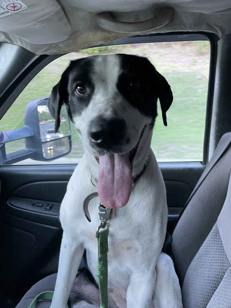
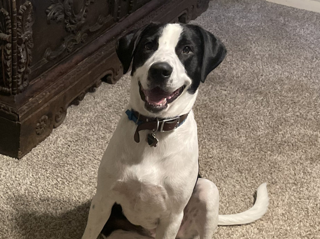
Like any other young dog, Delilah, a black Labrador Retriever-Great Pyrenees mix, loves to run and play. She wouldn’t be able to, however, if not for the knee replacement surgery she received at the Texas A&M School of Veterinary Medicine & Biomedical Sciences’ (VMBS) Small Animal Teaching Hospital (SATH).
Although knee replacements have existed in veterinary medicine for more than a decade, they’re fairly rare procedures because of the strict eligibility requirements. In fact, when Delilah had her surgery in March 2022, she became the SATH’s first knee replacement patient.
Just ten months later, Delilah’s artificial knee works just as it’s supposed to, allowing her to run and play to her heart’s content, much to the delight of her surgical team and her owner.
Discovering The Problem
Like all dog lovers, Delilah’s owner, Eric Golestan, was heartbroken when his time with his beloved first dog Huckleberry came to an end.
“I’d had Huckleberry for 12 years. He was born into my hand and I was really emotionally connected to him,” the Dallas native said. “When he passed away at the end of 2020, my mom thought I was depressed and found somebody who had a pregnant dog; one of the puppies ended up looking just like Huckleberry.”
When Golestan went to meet the Huckleberry lookalike, he fell in love with another puppy as well and ended up adopting both, soon to be known as Sampson and Delilah.
Only a few months later, Golestan noticed the first signs that Delilah was experiencing some pain.
“She was just running through the backyard and all of a sudden she let out a yelp and was holding up her left hind leg,” he said. “She immediately recovered and was running around like nothing was wrong, so I just thought she had bumped into something. I never really thought it was serious but it flared up again later. We took her to a local vet, who took some x-rays and found out it was osteochondrosis.”
Specifically, Delilah has osteochondrosis (OC), a developmental skeletal disorder in which improper cartilage development causes sections of cartilage to loosen and break off within joints like the knee, elbow, ankle, and shoulder. Although Delilah had signs of OC in both of her knees, only the left was damaged enough to cause her pain.
“At a very young age, she was missing a major component of the weight-bearing surface of the knee, a critically important joint for canine mobility,” said Dr. Brian Saunders, a VMBS associate professor and veterinary orthopedic surgeon. “Our normal treatment methods to address OC (such as arthroscopic surgery, followed by rest, rehabilitation, and medications) were not going to be able to address Delilah’s defect because it was so large.”
Because Delilah’s left knee was already so damaged, her activity had to be severely restricted; she was not allowed outside off-leash and wasn’t able to run and play with her brother—a serious hindrance for a puppy.
Feeling that Delilah’s quality of life was at stake, Golestan and Saunders began to discuss the alternative treatment option, a knee replacement surgery.
Weighing The Risks
For many dogs, knee replacement surgery is too risky to attempt.
“A lot of dogs with severe, end-stage arthritis of the knee have undergone many surgeries; in many of these cases, there is a documented or suspected infection at some point along the way,” Saunders said. “When there has been an infection in a joint, even if the infection is clinically resolved, there’s a high likelihood that a knee replacement will get infected, even if advanced measures are taken in surgery to prevent infection of the implants.”
The most common joint replacement in small animal orthopedics is the hip joint, because hip dysplasia is so common and because there is a successful “exit strategy,” or alternative plan, if a major problem were to develop with the implants, like an infection.
“If you do a knee replacement and something goes wrong, the stakes are much higher,” Saunders said. “The exit strategy is not nearly as appealing as it is for the hip; you have to either amputate the leg, fuse the knee in a standing position, or design and manufacture custom implants that work much differently than typical knee replacement systems.”
Since avoiding post-surgery issues is important, the majority of patients with knee problems are not eligible for a replacement because of their history of previous infections. But because Delilah was so young—only a year old by the time she arrived at the SATH—she had never had knee surgery or any other major medical issues, making her a candidate for knee replacement surgery.
After doing several x-rays, a CT scan, and health screenings, Saunders confirmed that Delilah was a good candidate for the procedure.
“On the preoperative side, the last thing we did was have a series of conversations with Mr. Golestan,” he said. “This is a lifelong investment in a pet, and a lot of long-term monitoring and follow-up care is necessary to make sure everything’s going well.”
Despite knowing the challenges of the procedure, Golestan decided it would be worth it.
“Because the OC was present in both legs, I was really worried that if her left knee was bad, she was going to compensate with the right, which would then cause the right to blow out eventually,” Golestan said. “I did not want to see her limping for the rest of her life.”
A Life-changing Solution
Prior to performing Delilah’s knee replacement, Saunders consulted with fellow orthopedic surgeon Dr. Jon Dyce, at Ohio State University, and used Delilah’s CT scan to make 3D-printed bone models of her femur (thigh-bone) and tibia (shin-bone). He also performed a number of practice surgeries to ensure the surgical team had the best chance of achieving success.
During Delilah’s surgery, Saunders used intra-operative tools called cutting guides to remove the cartilage from the femur and tibia. Next, metal implants were placed on the bottom of the femur and the top of the tibia, with a polyethylene (surgical plastic) liner inserted between them. These implants are somewhat shaped like a normal knee and allow the joint to function properly.
“The surface of these implants is intentionally made to resemble bone,” Saunders said. “The surface tricks the body’s bone into growing into the implant. Once the bone grows into it, it’s locked into place.”
After Delilah’s three-hour procedure ended, the real work began for Golestan.
“The post-operative aftercare instructions are pretty intense for the clients,” Saunders said. “A thumbnail sketch of the instructions is leash walks only for three months, no off-leash activity indoors or outdoors, two to four medications for several weeks after surgery, a fair amount of rehabilitation exercise, and a number of re-check visits for examinations and x-rays.”
Golestan rose to the challenge and credits his mom’s willingness to help care for Delilah with getting them through the recovery period.
It may have been tough, but it was worth it; at Delilah’s six-month post-surgery appointment, her implant was found to be fully secure and she was given the “all clear” to return to full activity levels.
“She runs at 110% and now she can do anything she wants,” Golestan said. “Delilah is the most intense dog I’ve ever known and if she can go through the recovery process and be successful, any dog can do it.”
The Benefits Of New Procedures
The SATH’s Orthopedics Service is now performing knee, ankle, and elbow replacements in addition to total hip replacements.
In addition to helping the canine patient, one of the benefits of these newer joint replacement procedures is the increased learning opportunities for SATH students, interns, and residents.
“It gives the students exposure to an advanced orthopedic procedure they normally wouldn’t see,” Saunders said. “Also, there are more training opportunities for the surgery residents who are on their way to becoming board-certified surgeons. We perform a number of advanced orthopedic procedures here, but to be able to add some of these other joint replacements to their training experience is a big step forward, in addition to being a big step forward for the small animal patients who benefit from the expertise provided by the VMBS’ orthopedic surgery team.”
###
For more information about the Texas A&M College of Veterinary Medicine & Biomedical Sciences, please visit our website at vetmed.tamu.edu or join us on Facebook, Instagram, and X.
Contact Information: Jennifer Gauntt, Director of VMBS Communications, Texas A&M College of Veterinary Medicine & Biomedical Sciences, jgauntt@cvm.tamu.edu, 979-862-4216

