
Texas A&M University Research Aims To Improve Lyme Disease Diagnostics
Story by Rachel Knight, VMBS Communications
Lyme disease, the fastest growing vector-borne illness in the U.S., according to the Bay Area Lyme Foundation, is challenging to diagnose and can only be treated in the early stages of infection. Once the infection spreads to the nervous and muscular systems, it is both harder to detect and less susceptible to antibiotics.
Research by two Texas A&M University scientists, however, is focused on improving Lyme disease treatment outcomes by developing a test that’s both more accurate and more efficient than the current test for the infection.
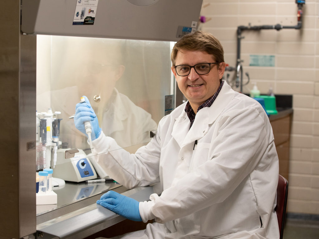
Dr. Artem Rogovskyy, an associate professor at the Texas A&M School of Veterinary Medicine & Biomedical Sciences (VMBS), and Dr. Dmitry Kurouski, an assistant professor in the Texas A&M Department of Biochemistry & Biophysics and the Department of Biomedical Engineering, are testing Raman spectroscopy, a technique used to detect vibrations at the molecular level, as a diagnostic tool for Lyme disease.
The results of Rogovskyy and Kurouski’s second paper on Raman spectroscopy as a diagnostic tool for Lyme disease, published in October, demonstrate that blood samples from mice and humans infected with the Lyme pathogen were more accurately identified with the Raman spectroscopy test than with the two-tiered serology, the only diagnostic method currently approved to diagnose Lyme disease in humans in the United States.
“We’re trying to develop a better test that would be simple, inexpensive, and accurate,” Rogovskyy explained. “By accurate, I mean highly sensitive and highly specific at the same time.”
The increased accuracy of Raman spectroscopy testing could improve Lyme disease diagnostic practices for both humans and animals believed to have been in contact with the disease.
For animals, the new test would require a smaller sample that could easily be taken in the field away from a veterinary clinic or hospital, thus improving mobile veterinary practices.
For humans, Raman spectroscopy testing could significantly decrease the amount of time needed to complete testing, increase the accuracy of the diagnosis, lower the cost of diagnosing the disease, and improve overall health outcomes by definitively diagnosing the disease earlier.
Rogovskyy said the team is in the process of validating the test through additional studies, and if the test is validated, it could become a very important tool for diagnosing Lyme disease worldwide, especially in more remote areas outside of the U.S. where the disease is prevalent, by enabling testing outside of traditional medical and hospital settings.
The researchers’ collaborative efforts have received funding from the Bay Area Lyme Foundation, a nonprofit that collaborates with world-class scientists and institutions to accelerate medical breakthroughs for Lyme disease. They also received human blood samples from the Lyme Disease Biobank, a clinical specimen repository.
Rogovskyy and Kurouski’s first paper published on Raman spectroscopy is the first proof-of-concept study to have explored Raman spectroscopy to diagnose mice infected with the Lyme pathogen. Their second paper included data on testing Raman spectroscopy on samples from mice infected with European Lyme pathogens, and also involved numerous human blood samples supplied by the Lyme Disease Biobank.
Rogovskyy anticipates the team may be able to publish more findings in about two years from the next phase of their research that entails testing human samples in a blind manner.
###
For more information about the Texas A&M College of Veterinary Medicine & Biomedical Sciences, please visit our website at vetmed.tamu.edu or join us on Facebook, Instagram, and X.
Contact Information: Jennifer Gauntt, Director of VMBS Communications, Texas A&M College of Veterinary Medicine & Biomedical Sciences, jgauntt@cvm.tamu.edu, 979-862-4216

