The most characteristic feature of domestic animals is their tame behavior.
This illustration, by Fabian Sinzinger, highlights the striking phenotypic differences between domestic and wild rabbits including changes in the size of the amygdala and the medial prefrontal cortex. After examining brain scans of domesticated rabbits, an international team of scientists has determined that domestication has had a profound effect on the morphology in particular regions of the brain.
Using high-resolution magnetic resonance imaging (MRI), the team—led by Texas A&M College of Veterinary Medicine & Biomedical Sciences (CVM) professor Leif Andersson—has found that domesticated rabbits’ amygdala and medial prefrontal cortex, those regions of the brain involved in fear processing, have been particularly effected. The study has been published in the Proceedings of the National Academy of Sciences of the United States of America (PNAS)
In contrast to domestic rabbits, wild rabbits have a very strong flight response because they are hunted by eagles, hawks, foxes and humans and, therefore, must be very alert and reactive to survive in the wild.
“In fact, Charles Darwin wrote in ‘On the Origin of Species’ that ‘no animal is more difficult to tame than the young of the wild rabbit; scarcely any animal is tamer than the young of the tame rabbit,’” Andersson said. “There is no doubt that this type of differences in behavior between wild and domestic animals to a large extent are genetically determined.”
In the study, scientists raised eight domesticated and eight wild rabbits under very similar conditions to minimize changes due to environmental effects. The brain MRI data were interpreted with sophisticated image analysis in which the scientist carrying out the analysis was unaware of the status animals—that is, wild or domestic.
“We observed three profound differences between the brains of wild and domestic rabbits,” said Irene Brusini, first author and doctoral student at KTH Royal Institute of Technology in Stockholm. “Firstly, wild rabbits have a larger brain-to-body size ratio than domestic rabbits. Secondly, domestic rabbits have a reduced amygdala and an enlarged medial prefrontal cortex. Thirdly, we noticed a generalized reduction in white matter structure in domestic rabbits.”
“These differences in brain morphology make perfect sense in relation to the fact that domestic rabbits are less fearful and have an attenuated flight response compared with wild rabbits,” said Mats Fredrikson, a professor at Uppsala University and Karolinska Institute, both in Sweden, and one of the senior authors on the paper.
The results show that an area involved in sensing fear, the amygdala, is smaller in size, while an area controlling the response to fear, the medial prefrontal cortex, is larger in domestic rabbits.
“The reduced amount of white matter suggests that domestic rabbits have a compromised information processing, possibly explaining why they are more slow reacting and phlegmatic than their wild counterparts,” Fredrikson said.
This study follows a previous study in which the team reported that genetic differences between wild and domestic rabbits are particularly common in the vicinity of genes expressed during brain development, according to Miguel Carneiro, from the University of Porto’s CIBIO-InBIO, in Portugal, who is one of the leading authors on the paper.
No previous study on animal domestication has explored changes in brain morphology between wild and domestic animals in such depth as Andersson’s team has done in this study, said Andersson.
“When we initiated the study, the concern was that any changes may be too subtle to be noticeable with MRI, but that was clearly not the case as we noticed distinct changes,” he said. “This study is not only important for our understanding of animal domestication but also for the basic understanding how variation in brain morphology can impact a complex behavior like fear response.”
###
For more information about the Texas A&M College of Veterinary Medicine & Biomedical Sciences, please visit our website at vetmed.tamu.edu or join us on Facebook, Instagram, and Twitter.
Contact Information: Megan Palsa, Executive Director of Communications, Media & Public Relations, Texas A&M College of Veterinary Medicine & Biomedical Science; mpalsa@cvm.tamu.edu; 979-862-4216; 979-421-3121 (cell)
Heartworms, which are spread by mosquitos, can cause serious health complications in pets. Although heartworms are more commonly diagnosed in dogs, cats are also at risk for this disease.
Megan Arroyo, a veterinary student at the Texas A&M College of Veterinary Medicine & Biomedical Sciences, explained how heartworms can harm our feline friends.
“The adult worms like to live in the right side of the heart and in the pulmonary artery, which is the blood vessel that supplies blood to the lungs,” Arroyo said.
“Adult heartworms can cause chronic inflammation that can cause permanent changes to the heart, and even heart failure.”
These symptoms, however, most commonly happen in dogs. Cats’ immune systems are better at fending off the worms before they become adults. It takes approximately six months for heartworms to become an adult in a new host, according to the American Heartworm Society.
However, fighting off heartworms can cause inflammation, which can lead to an immune reaction in cats. This syndrome, called Heartworm Associated Respiratory Disease, is associated with significant inflammation and permanent damage to the lungs, Arroyo said.
To protect your cat, you should give your pet preventative medication against heartworms, even if your cat primarily resides indoors.
“As much as we hate it, mosquitos definitely do get in the house, and it only takes one bite from one heartworm-infected mosquito for a cat or dog to get heartworms,” Arroyo said. “Heartworm disease in cats can cause irreversible lung damage, coughing, difficulty breathing, weight loss, and even sudden death. Why wouldn’t you choose to protect your beloved cat from this easily preventable disease?”
Prevention, Arroyo said, is key in protecting cats against heartworms because the treatments available for dogs are toxic to cats.
“If your cat has heartworms, all you can do is prevent further infection and manage symptoms,” Arroyo said. “Prevention truly is the best and only medicine for heartworms in cats. No cat parent should skip out on protecting their cat against heartworms.”
After all, kitty hearts deserve just as much love and protection as their canine counterparts, Arroyo said.
Pet Talk is a service of the Texas A&M College of Veterinary Medicine & Biomedical Sciences. Stories can be viewed on the web. Suggestions for future topics may be directed to editor@cvm.tamu.edu.
For Klause — a 2-year-old red dachshund with an infectious personality—the road to recovery seemed out of sight after a mauling by an unknown animal left him almost unrecognizable.
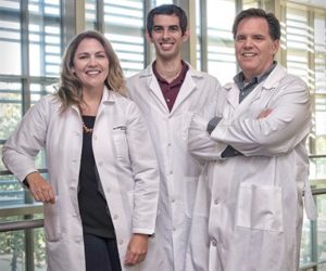
(From left) Cheryl Chadwick, Dr. Laurie Torkildsen, and Cassie Burghardt discuss Klause’s discharge after a mauling incident had left the dachshund with life-threatening injuries that require a pleurodesis procedure to heal.
But thanks to the work of a team of doctors at the Texas A&M Veterinary Medical Teaching Hospital (VMTH), Klause has returned home and is expected to make a full recovery after undergoing a rare pleurodesis procedure.
Klause’s owner Cheryl Chadwick had let Klause and his sibling Chloe out one last time before bed at around 10:30 p.m., when she heard a loud noise and raced outside to find the two. Seeing the extent of Klause’s injuries, Chadwick called Aggie veterinarian Dr. Kurt Kainer, who practices in Weimar, who told her she had two choices—leave Klause with him overnight, or bring him to Texas A&M.
When Klause arrived at the Small Animal Hospital (SAH), emergency room doctors found puncture wounds in his abdomen and a hole in his lung. Part of his liver also had been torn and was displaced in his abdomen.
Dr. Laurie Torkildsen, a third-year critical care resident at the SAH, said that Klause ranks in the top-5 most severe trauma cases of her career thus far.
Although the hospital deals with mauling incidents quite frequently, Torkildsen explained that the penetrating wounds to both Klause’s chest and abdominal cavity, as well as the damage to his lung, made this a rare and more severe case.
“Anytime we have any penetrating wounds into a body cavity, we flush it out so an infection doesn’t brew. Then we went into his chest. He was very critical under anesthesia and we almost lost him a few times,” Torkildsen said. “We were able to partially tie off his lung to try and stop the leaking, but he was not doing well enough for us to completely stop it.”
To complete the surgery, Torildsen performed a rare procedure called a pleurodesis, which requires taking blood and putting in into the chest cavity.
“The hope is that all of the things that make your blood clot will cause the hole to plug,” she said. “I actually used my own personal dog, took his blood and gave it to Klause. After the second procedure, it worked, and we were able to stop the leaking lung.”
Even after receiving a guarded prognosis and waiting through several difficult procedures, Chadwick said she never lost faith that her sweet Klause would return home.
“When they were going to start operating, they said there was a 50-50 chance of survival. Then they called me later on and said he wasn’t doing very well with the surgery, and I told them, ‘Just do what you’re going to do. He is in God’s hands and he is in your hands,’” Chadwick said. “I was not panicked about it. I just felt like he was going to be OK.”
That Chadwick was so calm about the ordeal may have been surprising, considering her bond with Klause. After not owning a pet since high school, the love and attachment Chadwick felt when she received Klause as a puppy certainly was a welcomed surprise for her.
“We named him Prince Klause, and then we added ‘von’ to his last name to make is sound more German, because of the dachshund’s German roots. His whole name is Prince Klause von Chadwick. It’s bigger than he is,” Chadwick joked.
“He instantly became mine. He lives in the house and he sleeps with me. None of my friends can believe it. They say, ‘Cheryl, what happened to you?’ she continued. “He has just really been life-changing.”
Chadwick also called upon her friends, family, and the total strangers who are part of an online dachshund community to pray for Klause’s recovery; not only did those strangers contact Chadwick and her daughter for daily updates, but they also contributed to his medical expenses.
Klause’s survival was just as important to Torkildsen and rest of the VMTH team.
Cassie Burghardt, a fourth-year veterinary student who cared for Klause tirelessly during his six-day stay, said Klause’s recovery was meaningful and rewarding in more ways than one.
“This would have been rewarding no matter what, but because this was my first surgery and really my first big case, to have him do so well and survive has been such a good experience for me,” Burghardt said. “Klause was the perfect patient. He’s the best guy, and he is so sweet, and his recovery has been amazing. I think we’ve all been saying that since the beginning.”
Chadwick, who was filled with emotion and gratitude for the VMTH when she arrived to pick up Klause, could not wait to take her little prince home.
“I feel that this is the premier veterinary hospital, internationally. It was a blessing for me to be able to get in my car, dead of the night, and bring him here,” she said. “I believe they were instrumental in saving his life.
“I almost wouldn’t change this experience. Had it not happened that night, it could have happened another night and rescue might not have been possible. Even though you could say, ‘Was there something I could have done differently?’ I think that no matter how bad, things always seem to go the way they’re supposed to,” Chadwick said.
“I thought he would be here another week. I thought I might not have the level of expertise needed to care for him in this condition, but the fact that they feel he is ready to go home makes me ecstatic. I am so excited.”
###
For more information about the Texas A&M College of Veterinary Medicine & Biomedical Sciences, please visit our website at vetmed.tamu.edu or join us on Facebook, Instagram, and Twitter.
Contact Information: Megan Palsa, Executive Director of Communications, Media & Public Relations, Texas A&M College of Veterinary Medicine & Biomedical Science; mpalsa@cvm.tamu.edu; 979-862-4216; 979-421-3121 (cell)
Veterinarians from the Texas A&M College of Veterinary Medicine & Biomedical Sciences (CVM) are seeking middle-aged, healthy dogs of any breed, or combination of breeds, to participate in a clinical trial as part of the Dog Aging Project’s second phase.
The growing focus of the aging research at Texas A&M—led by Dr. Kate Creevy, an associate professor in the CVM’s Small Animal Clinical Sciences Department—is to preserve function and extend a dog’s “healthspan,” rather than to treat age-related disease after it has already begun. “Healthspan” refers to the period of life when someone is active, healthy, and feeling good.
The second phase of Creevy’s project is a randomized, placebo-controlled, double-blind veterinary clinical trial in healthy, middle-aged dogs to test the hypothesis that the immunosuppressive drug rapamycin can improve age-related decline in cardiac function and increase healthspan in companion dogs.
Rapamycin has been approved by the United States Food and Drug Administration (FDA) for use in humans to prevent organ transplant rejection and certain forms of cancer. Rapamycin also has been studied in dogs with cancer as a chemotherapy drug.
In the study’s first phase, Creevy’s collaborative research team tested rapamycin in healthy dogs, finding evidence of mild improvements in the heart functions of those healthy dogs, with no significant side effects or adverse events.
Dogs enrolled in the second phase of Creevy’s study will receive rapamycin or a placebo three times a week for six months. They will have three follow up appointments throughout the course of one year and owners will be asked to complete regular surveys about their dog as the trial progresses.
To participate, dogs must be 6-10 years of age and weigh 40-80 pounds. At enrollment, dogs will receive a full physical exam, a full blood and urinalysis panel, a heartworm test, an echocardiogram, and participate in a cognitive assessment—all at no cost to pet owners.
For Michael Barnett, a member of the Fightin’ Texas Aggie class of ’81, Texas A&M University was a second home.
It was here that he watched his sister become a veterinarian, discovered his passion for landscaping, met his wife of 34 years, and sent his two daughters to college.
When Michael passed away in 2015, his family chose to honor him in a way that would have made him proud—by donating a bench and planting a tree outside of the Texas A&M College of Veterinary Medicine and Biomedical Sciences (CVM). The tree, a southern live oak, is located by parking lot 13, and the bench is located in the Veterinary & Biomedical Education Complex (VBEC) courtyard.
During his time as a student at Texas A&M, Michael worked as a lawn spray technician for A-Perm-O-Green Lawn. After graduating with a degree in accounting, Michael and his wife, Becky Barnett, purchased the company and his passion for people and the outdoors continued to flourish.
Becky describes her husband as a man with morals, character, and an infectious love for life.
“Mike never met a stranger, and he spoke to anyone,” she said. “Mike was always singing, whistling, or making corny jokes and was generally regarded as ‘the happiest man I ever met.’”
Michael carried the Aggie spirit with him wherever he went and loved the traditions and values that his beloved Texas A&M stood for.
“‘Howdy’ is how he addressed everyone, not ‘hi’ or ‘hello,’ but a big ole ‘HOWDY,’” Becky said. “Mike worked long, hard hours and was always on his feet. He generally did not like standing around. However, at Texas A&M football games, he did not take a seat. He wore his favorite A&M shirt and stood tall and proud as the 12th Man, and it was an honor for him to do so.”
When it came time for Michael and Becky’s daughters, Jennifer and Emily, to apply for college, they knew Texas A&M University was their first and only option.
Jennifer, ‘12, and Emily, ‘16, both received Bachelor of Science degrees in biomedical sciences. Jennifer is now a fourth-year veterinary student at Texas A&M.
“He always told our daughters that if they attended Texas A&M he would pay for their education, because ‘Texas A&M is the only university worthy of [his] hard-earned money,’” Becky joked.
Before Michael passed, he told Emily that he was working on a new song. The lyrics he had written were “sitting on a bench, counting the raindrops, thinking about you.” After Michael’s death, Emily finished writing the song for her father.
“The bench was donated by our family and relatives in his memory and it was perfect for the song lyrics he wrote the summer he passed away,” Becky said. “The tree was planted at the new vet school in memory of the Aggie ‘roots’ he established and for all the years he worked tirelessly doing what he loved—working outdoors and creating beautiful lawns and landscapes.
“There were so many places we could have chosen for the memorial, especially in the field he dedicated so much of his life to, but we just felt compelled that only one place fitting and deserving to honor his memory and the love he had for his children was Texas A&M vet school,” she said. “He was one proud Aggie dad.”
###
For more information about the Texas A&M College of Veterinary Medicine & Biomedical Sciences, please visit our website at vetmed.tamu.edu or join us on Facebook, Instagram, and X.
Contact Information: Jennifer Gauntt, Director of VMBS Communications, Texas A&M College of Veterinary Medicine & Biomedical Sciences, jgauntt@cvm.tamu.edu,979-862-4216
Summers are crucial for students, as evidenced by the internship, externship, technical, and professional development opportunities that fill students’ email inboxes and job boards in the months leading up to spring semester final exams.
Summer opportunities sometimes help students discover particular challenges of a specific field of work, while leading others to their dream job.
As part of a new program created by Dr. Dan Posey, academic coordinator for the Texas A&M Veterinary Medical Center (TVMC) at West Texas A&M University, and Dr. Dee Griffin, TVMC director, third-year veterinary students Pamela May and Michelle Morelli participated in a food animal production externship in West Texas. The program was developed for students who have completed their second year of veterinary school.
Posey tailored the program to fit the interests of program participants, but its goal is to expose students to the needs of rural West Texas food animal production.
May said the program revealed to her the stark contrast between veterinary practices in rural and urban areas.
“It’s very different in a rural area versus a teaching hospital or any of the private practices that are in Houston or Dallas,” May said. “It’s all very, very different. This program was a very big exposure to that.”
A South Padre Island native who is on the CVM’s food-animal track in order to pursue beef production medicine,
May did not grow up with a food animal background, but discovered a passion for food-animal medicine as Posey’s mentee, as well as through her experiences on the production tour and summer externship program Posey developed.
May began her summer working at a 50,000-head beef cattle feedlot and then finished with two-week rotations at mixed animal practices. She said her experiences exposed her to a different world of food animal production that she had yet to experience.
“Before I went on the production tour in May, I had never been to a feed yard, and it was different from what the media portrays a feed yard to be,” May said. “I got to learn how it works from the ground up. They taught me how to work cattle, a lot of the basics you don’t learn in school.”
Morelli’s experience was tailored to her interests in the dairy industry.
Growing up in the Philadelphia area of Pennsylvania, Morelli conducted small livestock projects in high school, but found her love for agriculture, and specifically dairy production, as an undergraduate at Penn State University.
Morelli spent three weeks at two different mixed-animal practices and rotated between two consultant veterinarians who have contracts with farms, mostly dairies. She said she experienced a wide range of cases and learned how to apply her classroom skills to real-life situations.
“At the mixed-animal practice, in the mornings, we would go to different dairy farms,” Morelli said. “In the afternoons, we would go back to the practice and we would see mostly small-animal patients. Sometimes farm animals would come in, so I got to see a lot of everything.”
Though their summer activities varied, May and Morelli both said the summer was invaluable to their education. Morelli said she faced challenges throughout the summer, but the growth she experienced made the challenges worth it. She encourages other students to not fear taking a risk.
“Getting up at 4 a.m. isn’t exactly the most fun, but putting yourself out there gets you a lot of really invaluable experience,” Morelli said. “There were a lot of things I did that I had never done before.
“I did a lot of first things. Was I necessarily ready to do them in that moment? No, but the veterinarians understood that and were there for me,” she said. “Taking those risks is what’s really important.”
May enjoyed the program location and hopes to return to the West Texas area to practice veterinary medicine.
“I do think that I will end up back there,” May said. “My main goal is making a difference in how producers view women in veterinary medicine, especially in food-animal medicine. There are a lot more women coming into the field, and I want producers to know that women can do what male veterinarians can do.”
As a student on track to become a food-animal veterinarian, Morelli said she hopes to repair the disconnect between consumers and producers in the conversation about food-animal production.
“I want to try and show people that the people who do produce animals that end up going to the food system really do care about the animals’ well-being,” Morelli said. “They don’t want to see their animals in pain or suffering, and they do everything they can to try and fix that. We all do care about the same thing.”
May and Morelli both said they could not have done this on their own, expressing gratitude toward Posey for all he did to create a program that enhanced their passion for the food-animal field.
“Dr. Posey made the experience what it was, and he made it perfect for me,” May said.
“It was great that Dr. Posey had the connections and set us up with the people who would go above and beyond for us,” Morelli said.
May and Morelli said Griffin, who also is a Texas A&M CVM clinical professor located at West Texas A&M, also played a large mentoring role for their respective programs.
Griffin was named mentor of the year for 2017 at the American Association of Bovine Practitioners (AABP) Conference in Omaha, Nebraska. May said the award is fitting.
“They started announcing the award by telling us to close our eyes and picture our mentor,” May said. “I pictured Dr. Posey and Dr. Griffin. Low and behold, Dr. Griffin was the one getting mentor of the year.”
Out of all the professional and technical development opportunities presented to veterinary students for the summer, May and Morelli encourage anyone to pursue the West Texas production program.
“It was an experience that I couldn’t get here (in College Station),” May said. “I would encourage anyone to pursue this program.”
“If you want to get a real, hands-on experience and figure out how things are done with clients and how to get through the decision-making process, this is a really great experience,” Morelli said.
###
For more information about the Texas A&M College of Veterinary Medicine & Biomedical Sciences, please visit our website at vetmed.tamu.edu or join us on Facebook, Instagram, and Twitter.
Contact Information: Megan Palsa, Executive Director of Communications, Media & Public Relations, Texas A&M College of Veterinary Medicine & Biomedical Science; mpalsa@cvm.tamu.edu; 979-862-4216; 979-421-3121 (cell)
Many of us know someone affected by cancer. Unfortunately, cancer in the mammary glands (similar to breast cancer in humans) can also occur in both male and female dogs. In fact, mammary tumors are the most common type of tumor seen in intact (not spayed) female dogs.
Dr. Brandan Wustefeld-Janssens, assistant professor of surgical oncology at the Texas A&M College of Veterinary Medicine & Biomedical Sciences (CVM), described some of the clinical signs of mammary tumors in dogs.
“The most common clinical finding is a firm, non-painful nodule associated with the mammary chain,” Wustefeld-Janssens said. “These nodules can range from very small to very large and may grow fast. Continued growth of bigger tumors can lead to the skin overlying the tumor to become thinned, which can cause the skin to ulcerate, bleed, and become infected.”
Tumors may also be swollen, hot, and painful to the touch.
However, only about 50 percent of mammary tumors are cancerous, with a majority of these cases being carcinoma (arising from the lining of the ducts in the mammary gland), Wustefeld-Janssens said.
Luckily, you can take preventative action to help lower your pup’s risk of developing a mammary tumor.
Dr. Heather Wilson-Robles, associate professor of oncology at the CVM, said that spaying or neutering your pet before they have their first or second reproductive season is the most effective way of preventing mammary tumors in dogs. Regular checkups at the veterinarian is also a good way to protect your pet.
“Yearly physical examinations of both male and female animals should include careful palpation of the mammary chains,” Wilson-Robles said. “Any nodule should be further investigated; often, this would include some form of biopsy. We strongly recommend against a ‘wait-and-see’ approach—any mammary tumor that is growing rapidly or is swollen, hot, or painful should be seen by a veterinarian as soon as possible.”
The chances of a tumor being cancerous increases with age, so pay special attention to any abnormalities in your older pup’s mammary glands.
If your dog does develop a tumor, surgery is the standard of care for mammary tumors. In cases where there is high risk of tumors reoccurring or spreading, radiation and chemotherapy may be used for treatment.
We can’t control the development of cancer, but we can learn the signs and symptoms of the disease and report any abnormalities to a veterinarian. Additionally, pet owners can help prevent mammary tumors by considering spaying and neutering their dog.
Pet Talk is a service of the Texas A&M College of Veterinary Medicine & Biomedical Sciences. Stories can be viewed on the web. Suggestions for future topics may be directed to editor@cvm.tamu.edu.
Texas A&M’s GoWeb team has recognized the Center for Educational Technologies (CET) for its contributions to transformational learning at Texas A&M University with 2018 GoWeb Awards.
The center was presented the 2018 GoWeb Award for Transformational Learning, and CET senior IT professional III Dan Shuta received runner-up recognition for a 2018 GoWeb Award for Innovation for his creation of StepStone, a web-based elearning authoring tool application.
The awards were presented on June 8 during Texas A&M’s Annual GoWeb Retreat.
GoWeb is an inclusive, collaborative community of professionals dedicated to supporting one another and elevating web communication across campus. The GoWeb Awards recognize individuals and teams that create innovative web communications supporting the university’s three pillars: impact, innovation and transformational learning.
Comprising a multi-disciplinary team of 10 highly experienced faculty and multimedia production staff who produce a wide range of transformational learning experiences, the CET has exemplified excellence in the transformational learning occurring within the College of Veterinary Medicine & Biomedical Sciences (CVM) by championing mobile technologies, collaborating with units across the university, and creating educational solutions for faculty and students.
“The CET incorporates the latest in educational technologies to transform education for all students at Texas A&M,” said CET director Nicola L. Ritter. “These immersive educational experiences prepare faculty and students to address the world’s most formidable problems and selflessly-serve the world around them.”
Among the ways the center has embraced transformational learning is through its partnership with the DVM curriculum committee and faculty teaching in the DVM program to harnesses educational technologies to support the new curriculum, which recently completed its inaugural year.
The active, online learning communities for veterinary students and faculty has expanded the CVM’s culture of in-person collaborations to online collaborations, enabling CVM faculty to continue conversations on teaching and learning outside of meeting rooms and share new information more effectively around teaching and learning topics.
“The online learning community for students provides a one-stop-shop for all things related to the DVM program and their progress within the program,” Ritter said. “Students can communicate with their peers and instructors inside and outside of class more easily than ever before. With everything DVM in one place, program decision-makers can observe interactions from afar using the platforms robust analytics reporting capabilities that combine learning analytics and data visualization.”
The CET also has championed the veterinary program “going mobile” by creating device-agnostic, platform-independent and easy-to-use digital educational resources accessible to all. This includes the development of StepStone, a content authoring software that allows educators to create a variety of learning experiences accessible from any internet-enabled device.
“StepStone is a great example of leveraging educational innovations to support transformational learning,” Ritter said. “The CVM now has a scalable solution that allows them to rapidly produce e-learning materials and enhance every course in their new veterinary program.”
Through its collaboration with TAMU’s IT Accessibility and Disability Services, the CET developed an inclusive teaching and learning culture that includes training faculty on accessibility topics; creating accessible online instructional materials for students; and consulting with other units on campus to provide transformational learning experiences for all.
“The CET has embedded accessibility into every aspect of the unit, from creating an inclusive culture to applying universal design teaching techniques to adding robust accessibility testing,” Ritter said. “The CET continues to lead by example in transformational learning for all by developing innovative learning experiences and sharing the CET’s experiences with others.”
Shuta has created more than 150 technology applications for TAMU and worked with approximately 100 educators to make their visions become a reality. Approximately 10,000 students and 200 instructors have impacted by his work, according to Ritter.
“Dan is a brilliant programmer and talented illustrator at Texas A&M,” she said. “He has over 25 years of experience in web communications, specifically for the purpose of learning. He is a lifelong learner and thrives on creating educational innovations to share information with others in an engaging way.”
Among his contributions to transforming learning was his development of StepStone, which has been used by approximately 100 educators to created more than 200 online educational resources since its inception.
“Dan single-handedly developed a tool that allows TAMU to rapidly develop instructional materials. This has improved TAMU’s content development and others inside and outside the college have taken note,” Ritter said. “Under his leadership StepStone continues to grow and his efforts have aided in making this product sought after by other colleges at TAMU and worldwide. StepStone has also opened the door to other fields of collaboration.”
To ensure StepStone continues to meet the needs of its users, Shuta is collaborating with TAMU’s IT Accessibility and Disability Services to add accessible functionality to all the features available in StepStone.
To learn more about the center’s contributions to transformational learning at TAMU, visit the CET’s submitted portfolio at tamucet.org.
###
For more information about the Texas A&M College of Veterinary Medicine & Biomedical Sciences, please visit our website at vetmed.tamu.edu or join us on Facebook, Instagram, and Twitter.
Contact Information: Megan Palsa, Executive Director of Communications, Media & Public Relations, Texas A&M College of Veterinary Medicine & Biomedical Science; mpalsa@cvm.tamu.edu; 979-862-4216; 979-421-3121 (cell)
Feral cats may look cute and cuddly, but they are a lot different than a typical house cat.
While feral cats are considered wild animals and prefer to live their lives outdoors, their kittens can be rescued from the streets and put into loving homes as pets.
Feral cats have never been socialized to humans. In fact, feral cats are generally afraid of people and may even run away if approached, according to Megan Arroyo, a veterinary student at the Texas A&M College of Veterinary Medicine & Biomedical Sciences.
Because feral cats live their entire lives independent from humans, they may not be spayed or neutered by a veterinarian. This can lead to an overpopulation of feral cats, as well as kittens being born in unsafe areas.
However, local pet shelters, veterinarians, and other programs may be able to help prevent the overpopulation of feral cats by giving kittens born to feral cats a good home.
“If you find a potentially feral cat with a litter of kittens, one of the best places to start is by contacting your local animal shelter or animal control,” Arroyo said. “They should be able to help you to decide the most appropriate action to take, which is often trapping the mom and the kittens so that the mom can be spayed and the kittens socialized and placed in homes.”
A properly socialized kitten is exposed to humans and other species, such as dogs, between the ages of 2 to 7 weeks, Arroyo said. Once they are weaned from their mother, at around 4 to 6 weeks, kittens can socialize even more in a foster home or their permanent home.
“Socialization is basically what it sounds like—you want to expose the kitten to experiences you would like it to be comfortable with as an adult, such as gentle handling and petting,” Arroyo said.
Socialization is beneficial because it gives kittens a better chance at finding a loving home that can provide veterinary care, which can help significantly increase the kitten’s lifespan, Arroyo said.
Since adult feral cats prefer to live out their life in the wild, the mother feral cat will typically be re-released after she is spayed and her kittens are weaned.
“This allows her to live out her life where she is happiest, but keeps her from contributing to the feral cat population,” Arroyo said.
There are many benefits of rescuing kittens born to feral cats, but the biggest reward is knowing that you saved a pet’s life.
Pet Talk is a service of the Texas A&M College of Veterinary Medicine & Biomedical Sciences. Stories can be viewed on the web. Suggestions for future topics may be directed to editor@cvm.tamu.edu.
Dr. Ashley Saunders, Dr. Mark Wierzbicki, and Dr. Duncan Maitland
For decades, biomedical engineers have used their acumen to revolutionize healthcare through the development of devices, tools, equipment, techniques, and pharmaceuticals that have advanced the medical field in ways previously unimaginable.
While patients around the world have benefited from this ingenuity, those patients almost exclusively have had one thing in common—they’ve all been human.
Researchers in the Texas A&M College of Veterinary Medicine & Biomedical Sciences (CVM) and the College of Engineering have teamed up to begin filling that gap in the biomedical engineering field—that of veterinary medicine—by exploring the possibilities of what can be accomplished when innovative minds come together.
Getting to the Heart of the Problem
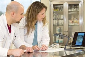
Saunders trains cardiology resident Derek Matthews using the model she helped develop.
Dr. Ashley Saunders, a professor of cardiology and clinician in the Veterinary Medical Teaching Hospital’s (VMTH) small animal cardiology service, began using 3-D imaging and printing to create models of the heart to aid in the teaching of anatomy and preparing for complex surgeries for animals. But while 3-D models are useful in this regard, the technology has not been a definitive training tool, especially in the case of treating minimally invasive cardiology defects.
Cardiology residents are taught heart and blood vessel catheterization through observation and practice, relying on an understanding of the anatomy and the feel of inserting a catheter to perform a procedure.
Teaching catheterization using 3-D printed models is difficult because doctors can’t see inside the blood vessels they’re trying to navigate, and it’s also difficult to replicate the feel of an animal’s blood vessels; therefore, doctors have to learn catheterization on a beating heart.
“That’s how you learn,” Saunders said. “That’s how I learned.”
Because heart defects like patent ductus arteriosus (PDA)—a congenital defect characterized by an opening between two blood vessels leading from the heart—are the most commonly addressed congenital defect by cardiologists at Texas A&M’s Small Animal Hospital (SAH), Saunders began looking to create a safer environment in which residents could learn and practice, one in which the stakes weren’t quite so high.
Enter Dr. Duncan Maitland, the Stewart & Stevenson Professor I in the College of Engineering’s Department of Biomedical Engineering, and Dr. Mark Wierzbicki, a post-doctoral researcher in Maitland’s Biomedical Device Laboratory.
Synthesizing a Solution
The model simulates a real PDA procedure by allowing students to feel what it’s like to pass a catheter while watching the movement on a screen.
Maitland, whose research focuses on novel treatments for cardiovascular disease, had previously worked with a VMTH cardiovascular doctor to create devices for use in the operating room; the doctor encouraged Maitland and the biomedical engineers in his lab to move toward creating devices for animal patients.
“I learned long ago that we could reduce the number of animal iterations on device development if we brought clinicians in, or even imported our models into the clinical environment for more advanced testing,” Maitland said. “Just getting iterations on models of real anatomies reduces the number of animals that need to be used in studies, and the quality of devices goes up exponentially.
Maitland’s lab had developed blocks made of silicone to test devices created to treat cardiovascular diseases. One day, Saunders toured Maitland’s lab and immediately began thinking about how the technology could be applied to help her train residents to treat PDA.
“The silicone block was made to resemble a PDA, and I knew we could use the 3-D prints from our patients to make one that is more anatomically correct,” Saunders said.
And that’s exactly what she and Wierzbicki, a doctoral student at the time, did. The two put their heads together and devised a plan that would combine the 3-D imaging technology Saunders was already using with the silicone-based technology Wierzbicki was exploring for his dissertation.
The final product looks like a clear, rubber block, inside which, upon closer inspection, has the outline of several “tubes.” These tubes are arteries cast from the actual heart from one of Saunders’ canine patients. To make the cast, Saunders used CT scans to create a 3-D representation of the dog’s heart printed on the 3-D printer in Maitland’s laboratory.
“We were able to 3-D print the CT scanned heart using a dissolvable material and vapor polish the printed model to smooth out the ridges from the 3-D printing process,” Wierzbicki said. “We took the smooth, 3-D printed heart, cast silicone around the model, and then dissolved out the 3-D-printed part. After completing those steps, we were left with a model Dr. Saunders could use for training.”
The result was a solution to multiple problems—not only did the project become part of Wierzbicki’s dissertation, but it produced anatomically correct, customized models that might change the way budding cardiologists are trained to learn catheterization techniques and repair heart defects.
To create an even more realistic setting, Saunders incorporated a camera that projects what the doctors are seeing onto a computer screen so that they train on a simulated heart that mimics a true procedure.
“When we do these procedures in a dog, we can’t see inside the body; we use fluoroscopy, with images displayed on a screen that we have to look up at. So, it is important to learn how to do these procedures by watching a screen,” she said. “We can mimic the procedure by having the silicone blocks, because they have the anatomy inside, and the block is clear, so they can see through it; the document camera displays the image up on my computer screen.
“They watch as they pass a catheter in and they learn how to do the procedure by getting the feel of inserting a device,” she said. “It doesn’t require fluoroscopy or radiation, and it doesn’t require them being inside an actual dog to practice for the first time.”
The best part—the silicone blocks are virtually indestructible. “This means you can take the block into a training lab setting, knowing that it’s going to stand up to being used over and over again,” Saunders said.
The Future of Biomedical Engineering: Just a Heartbeat Away
Saunders, Maitland, and Wierzbicki
Because of what the model means for how doctors treat cardiac defects, Saunders, Wierzbicki, and Maitland have published multiple papers related to the model and other devices used in cardiology.
Saunders also has begun using the model in training exercises and labs for both residents and specialists who have an interest in catheterization techniques and interventional cardiology. During a recent training that included specialists from around the world, Saunders found that the doctors responded enthusiastically to the model.
“They really loved it,” she said. “They said they feel like they’re more comfortable practicing with the model; it makes more sense to them.”
While most companies that manufacture devices for human cardiology currently aren’t interested in making devices for animals, as more and more veterinary surgeons begin recognizing the value of this kind of model, Maitland said he hopes that attention will open the doors for more opportunities for collaboration between biomedical engineers and the field of veterinary medicine.
“If you look at all of engineering, and biomedical engineering, specifically, you don’t think about animal health care as a primary focus. We’re not trained to do that, and so there are not enough partnerships going on between the two colleges, and specifically biomedical and the CVM,” said Maitland, who is chairing a committee in his department to do just that. “I think we could make a lot of impact, and in this case, not just with what Ashley is developing for training, but we can also impact the technologies that are used in animal health care significantly, if we just pay attention to it.”
###
For more information about the Texas A&M College of Veterinary Medicine & Biomedical Sciences, please visit our website at vetmed.tamu.edu or join us on Facebook, Instagram, and Twitter.
Contact Information: Megan Palsa, Executive Director of Communications, Media & Public Relations, Texas A&M College of Veterinary Medicine & Biomedical Science; mpalsa@cvm.tamu.edu; 979-862-4216; 979-421-3121 (cell)


 Heartworms, which are spread by mosquitos, can cause serious health complications in pets. Although heartworms are more commonly diagnosed in dogs, cats are also at risk for this disease.


 Many of us know someone affected by cancer. Unfortunately, cancer in the mammary glands (similar to breast cancer in humans) can also occur in both male and female dogs. In fact, mammary tumors are the most common type of tumor seen in intact (not spayed) female dogs.
 Texas A&M’s GoWeb team has recognized the Center for Educational Technologies (CET) for its contributions to transformational learning at Texas A&M University with 2018 GoWeb Awards.