Texas A&M Superfund Researchers Establish Green Infrastructure Plan For Houston Community

A graphic of the master plan for sunnyside, showing a lot of green spaces
Participatory master plan and schematics developed for Sunnyside, Houston, TX, USA.

Texas A&M Superfund Research Center investigators have developed a novel green infrastructure plan to reduce stormwater runoff in Houston’s Sunnyside community by uniquely combining the results of three separate landscape performance tools.

Green infrastructure (GI) is a network of interconnected green spaces that reduce the impact of flooding in urban areas. In communities like Sunnyside, which has 14 facilities within a mile that release toxins into the air and water, GI can also reduce the amount of contaminants in stormwater runoff.

Several tools have been created to measure the impact of GI, flooding, and runoff, but by combining these tools in a way never done before, Superfund researchers were able to put together a plan that would not only reduce flooding immediately but also create a healthier and more sustainable community for years to come.

“Sunnyside has a lot of vacant properties but it doesn’t have a lot of money to build things like levies. The idea was that we would use green infrastructure as a cheaper way to help combat a lot of the runoff and flooding issues,” said Dr. Galen Newman, principal investigator of the Superfund Center’s Community Engagement Core.

“The longer GI stays in place, the more effective it is over time,” he said. “We wanted to show Sunnyside that if they invest a little up front, it will pay for itself in the long run.”

Why Sunnyside?

Newman, who is also an associate professor in Texas A&M’s Department of Landscape Architecture and Urban Planning, worked with the non-profit organization Charity Productions to choose Sunnyside as the project site.

“We really wanted to target underserved communities that have both flood issues and contamination issues,” Newman said. “We did a lot of mapping of previous floods and the interesting thing about Sunnyside is that even though it’s on the Sims Bayou, most of what flooded was the ‘ponding,’ or standing water, areas.”

Sunnyside, which is one of Houston’s oldest historically Black communities, has been hit hard by many of the recent hurricanes that have struck along the Gulf of Mexico. A 2019 survey found that one-third of the homes in Sunnyside were damaged by recent storms and that poor drainage/flooding is the top problem according to community residents.

9 graphics showing different characteristics of the Sunnyside site, such as percent impervious surface and number of parks
The site’s spatial characteristics and conditions in Sunnyside, Houston, TX, USA.

Sunnyside is also considered a “fenceline” community because it is immediately adjacent to an industrial center and is directly affected by the chemical emissions or other operations of the company.

“If a neighborhood is tangent to a lot of industrial contaminants and it gets flooded, all of those contaminants wash into the community,” Newman said. “Some of the neighborhoods we work with that are in close proximity to these industries have pretty elevated cancer, obesity, and high-cholesterol rates. If you could have specific areas to store and clean those flood waters, you’re not going to get as many public health issues from being exposed to that contamination in the long term.”

One of Sunnyside’s largest health concerns is the prevalence of asthma, especially when compared to the surrounding areas.

The Houston Health Department found that the percentage of residents with chronic asthma was more than twice as high in Sunnyside as in the rest of Harris County (the county containing Houston), and that 14.3% of children in Sunnyside have asthma, compared to 8.9% in the county as a whole.

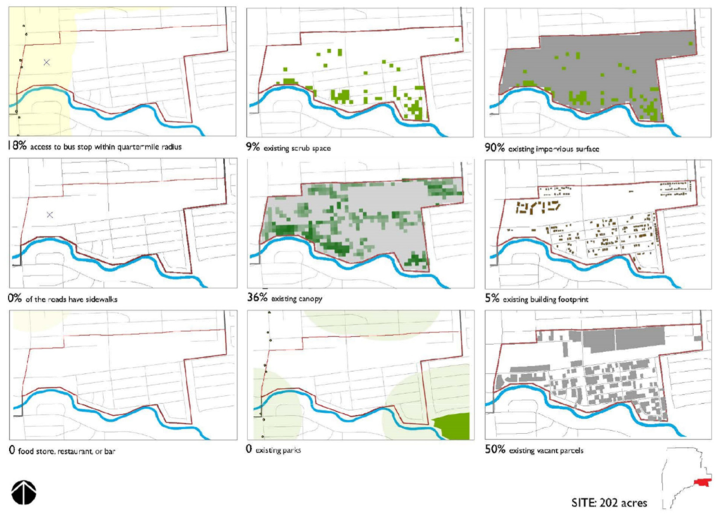
Recognizing the importance of addressing the flooding and public health issues in Sunnyside, community members worked directly with the Aggie researchers during a series of meetings and tours to select a 202-acre area within the community as their project site.

This collaboration with community members did not stop once the site was selected, however; resident input was an important part of the project from beginning to end.

“The community members know more about what’s going on than we ever will, so getting their feedback is key,” Newman said. “It’s super important to have them involved throughout. They can provide ground truthing (the process of checking the results of machine learning for real-world accuracy) for our data and help us propose solutions that will not only work but also fit with how the community wants to grow.”

Residents’ involvement also helps make sure that the final plan is one that can actually be implemented, in regards to regular maintenance and cost.

Developing The Plan

By combining the results from three separate landscape performance tools that quantify different aspects of GI, the researchers were able to get a complete picture of what was needed to reduce runoff and flooding in Sunnyside.

“There are about six applicable performance tools that we can mix and match in different ways. In this case we used three, which is probably the most ever combined for one project,” Newman said. “By mixing tools, you can show much broader impacts. In this case, we were able to combine them to show the runoff reduction, the contaminant load decrease, and the economic benefit of green infrastructure.”

The three tools used were the Value of Green Infrastructure Tool (VGI), which assigns economic value to GI practices and investments; the Green Values National Stormwater Calculator (GVC), which assesses the effectiveness and cost of stormwater management practices; and the Long-Term Hydrologic Impact Assessment Model (L-THIA), which estimates long-term runoff and the amount of pollution it contains.

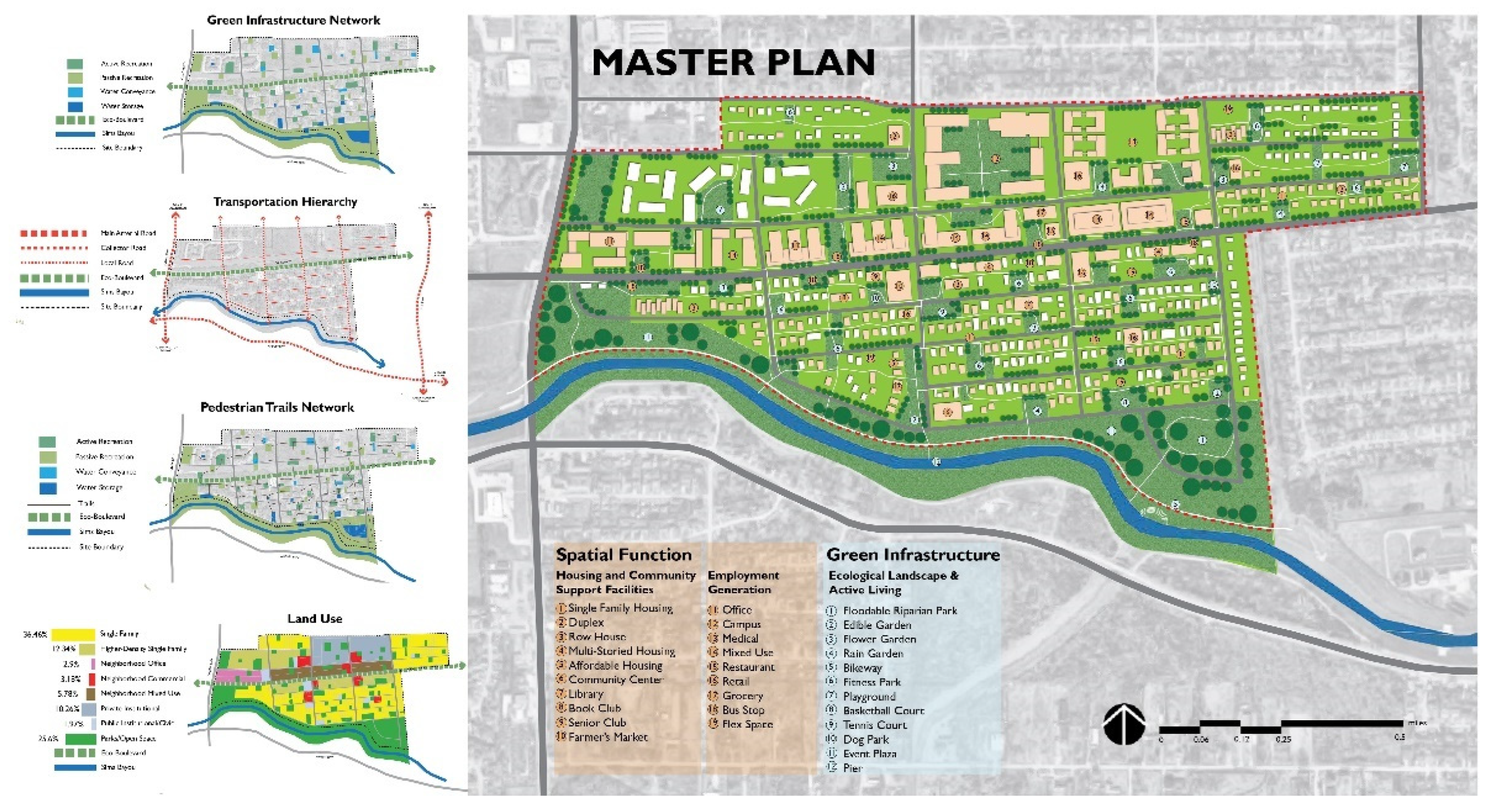
Newman, other Superfund researchers, and Texas A&M students spent a year and a half collecting the raw data and turning it into an actionable plan that not only reduces runoff and contaminants but also creates new recreational spaces, housing, and job opportunities for the community.

“The issue is that there’s so much impervious surface (such as roads and parking lots) because we develop so rapidly and haphazardly. Water doesn’t have anywhere to go; it’s just accumulating,” Newman said. “Our approach breaks up a lot of the monotony of the impervious surfaces and gives the water somewhere to be stored.”

At the same time, the plants added to the area pull contaminants from the water through a process known as phytoremediation.

“Green infrastructure—from bioswales and rain gardens to larger things like vegetated, riparian areas and constructed wetlands—not only slows the runoff volume down to the degree where it’s naturally phytoremediated but it also allows it much more time to seep down into the aquifer,” Newman said.

Recharging aquifers supplies the community with more clean water in the long-term, providing benefits for years to come.

Once their work was done, the researchers provided Sunnyside residents with a final proposal containing the details of the plan, its many benefits, and possible funding options. Now, it’s up to the community members to make it a reality.

Find the full publication at https://www.mdpi.com/2071-1050/14/7/4247/htm

###

For more information about the Texas A&M College of Veterinary Medicine & Biomedical Sciences, please visit our website at vetmed.tamu.edu or join us on FacebookInstagram, and X.

Contact Information: Jennifer Gauntt, Director of VMBS Communications, Texas A&M College of Veterinary Medicine & Biomedical Sciences, jgauntt@cvm.tamu.edu, 979-862-4216

CVMBS Faculty Listed Among Top 2% Of Scientists

the VENI building

Sixteen faculty members from the Texas A&M College of Veterinary Medicine & Biomedical Sciences (CVMBS) have been listed among the top 2% of scientists in the world, according to Stanford University’s list of the most cited scientists.

Dr. Stephen Safe, distinguished professor of toxicology in the CVMBS Department of Veterinary Physiology and Pharmacology (VTPP), was the highest listed CVMBS faculty member and the second highest faculty member at Texas A&M University. His ranking of 488 puts him in the top .01% worldwide.

Safe’s career has focused on researching medical compounds to treat and cure cancers; he was inducted as a fellow by the American Association for the Advancement of Science (AAAS) earlier this year and his research has been cited more than 43,000 times since 1996.

Dr. Michael Criscitiello, CVMBS associate dean for research and graduate studies, said having this many faculty on the list demonstrates the quality of research in the CVMBS and the impact CVMBS research has beyond College Station and Texas.

 “Our faculty members are working across disciplines at Texas A&M and with federal and industry partners to develop innovative solutions to problems that challenge the world today,” said Criscitiello. “As importantly, these CVMBS faculty scientists are also inspiring the next generation of researchers through education and mentorship, ensuring that Aggies will continue to play a critical role in protecting animal, human, and environmental health for decades to come.”

The list also includes at least one faculty member from every CVMBS department, which Dr. John August, the Carl B. King Dean of Veterinary Medicine at Texas A&M University, said shows the breadth and scope of the quality work being done within the college.

“Each day, through clinical trials in our Veterinary Medical Teaching Hospital and innovative and translational scientific research, faculty members in the College of Veterinary Medicine & Biomedical Sciences are making discoveries that will improve the lives of animals and human beings,” August said. “Embracing the Aggie core values of excellence, service, and leadership, these highly accomplished scientists are advancing both veterinary and human medicine so that doctors around the world can continue to offer the most advanced patient care.”

These rankings are based on data from Elsevier’s Scopus, an abstract and citation database; the list comprises more than 186,000 scientists from universities, hospitals, institutes, industries, and laboratories around the work working in 22 scientific fields and 176 subfields. Rankings are measured by a combination of c-score (a metric based on citations to published papers), h-index (which measures productivity and citation impact of publications), and other factors.

Along with Safe, other current CVMBS faculty on the list include:

In addition, CVMBS retired faculty listed for their career-wide accomplishments include:

  • Friedhelm Schroeder, senior professor, Department of Veterinary Physiology & Pharmacology
  • Larry Johnson, professor, Department of Veterinary Integrative Biosciences
  • James Womack, senior professor, Department of Veterinary Pathobiology
  • Katrin Hinrichs, professor, Department of Veterinary Physiology & Pharmacology
  • Ian Tizard, professor, Department of Veterinary Pathobiology
  • Michael Willard, senior professor, Department of Small Animal Clinical Sciences
  • Joe Kornegay, professor, Department of Veterinary Integrative Biosciences
  • R.R. Pool, clinical professor, Department of Veterinary Pathobiology
  • Steven Brinsko, professor, Department of Large Animal Clinical Sciences
  • Thomas Craig, professor, Department of Veterinary Pathobiology
  • George Lees, professor, Department of Small Animal Clinical Sciences
  • John Edwards, professor, Department of Veterinary Pathobiology
  • J.B. Puschett, research professor, Department of Veterinary Pathobiology
  • T. Tiffany-Castiglione, professor, Department of Veterinary Integrative Biosciences

###

For more information about the Texas A&M College of Veterinary Medicine & Biomedical Sciences, please visit our website at vetmed.tamu.edu or join us on FacebookInstagram, and X.

Contact Information: Jennifer Gauntt, Director of VMBS Communications, Texas A&M College of Veterinary Medicine & Biomedical Sciences, jgauntt@cvm.tamu.edu, 979-862-4216

A ‘Productive’ Endeavor: Veterinary Research In The Texas Panhandle

The VERO building

Identifying and solving problems that challenge the veterinary profession often require a team effort, which is why researchers in the Texas A&M School of Veterinary Medicine & Biomedical Sciences’ (SVMBS) Veterinary Education, Research, & Outreach (VERO) initiative are working in partnership with veterinary practitioners, animal scientists across the country, and the livestock industries to address issues that will make a significant difference in animal health and welfare.

“We view veterinarians as key partners in the overall mission to improve the health and well-being of livestock,” said Dr. Paul Morley, director of research at VERO. “A significant part of identifying the health issues that we address stems from veterinarians’ unique roles in the oversight and championing of the health and welfare of animals.”

Since Morley joined VERO in 2019, he has been a big champion of fulfilling the SVMBS’ mission of “Serving Every Texan Every Day”—which is also central to Texas A&M University’s mission as a land-grant university.

Key to that has been developing a research program that strives to be productive, impactful, and collaborative. And in just three short years, VERO’s research has had success in all three of those dimensions.

“It’s pretty remarkable that since September 2020, when we moved into the new VERO building, we went from four faculty and one staff member to 21 faculty and over a dozen staff, including the two new faculty we hired on the research team in 2021, Dr. Robert Valeris-Chacin and Dr. Matthew Scott,” Morley said. “The entire team has been highly successful, as shown by our active grants in 2021 and 2022, which total $4.9 million. In addition, since 2019, VERO faculty have published 53 peer-reviewed publications.

“While there are four faculty who have a predominantly research-associated appointment, all of the faculty working at VERO have scholarly activity as part of their roles,” he said. “So the entire VERO team is contributing to the research mission.”

Solving Problems Through Discovery

As an epidemiologist whose research interests have always been driven by the livestock industry’s needs, Morley sees the primary goal of VERO scientists as pursuing research that will improve the prosperity of the Texas Panhandle’s rural communities and livestock industries; they do that, he says, by examining issues that advance animal, human, and environmental health and that are important on all levels—locally, nationally, and globally.

“We strive to make a global impact, to translate our discoveries into products of knowledge and innovation,” Morley said.

As such, the VERO research team is tackling a wide range of topics, including respiratory and gastrointestinal health, microbial ecology, antimicrobial resistance, food safety, genomics and transcriptomics, lameness, and reproduction.

Of particular interest—both to VERO researchers and the livestock industries—is bovine respiratory disease (BRD), a problem that is critical to Texas agriculture and the state at large. Because BRD is both a health and an economic problem within cattle production, six of the VERO team’s nine United States Department of Agriculture (USDA)-funded studies focus on that issue.

In another federally funded project, the Food and Drug Administration issued a call for grants to address the problem of liver abscesses in cattle, and the Texas Cattle Feeders Association approached SVMBS scientists to lead in writing this grant, which was supported by representatives from the three largest cattle feeding companies in the world and the largest veterinary consulting groups.

That $500,000 grant led to a trial in which VERO researchers—working with livestock producers—are examining whether using lower amounts of antibiotics can be equally effective in preventing liver abscesses. The project is being conducted in partnership with faculty at West Texas A&M University (WT), Colorado State University, and Michigan State University.

“The Food and Drug Administration, and the public, would like us to use fewer antimicrobial drugs, and the industry is interested in doing that,” Morley said. “This example highlights the successes of how the industry came to us with a problem and asked us to help, and then we worked as an integrated team to address the issue.”  

Also important to Morley is that VERO researchers employ cutting-edge technology such as molecular biology and super computers to study the genomics and gene expression of animals and their microbiome.

“We’re looking at the overall health of animals and approaching long-standing, important problems in really novel ways. For example, we are using state-of-the-art sequencing technology to investigate the entire microbial community, the microbiome, in relation to infectious diseases by focusing on one particular bacterium,” Morley said. “By using these cutting-edge techniques, we have new insight on problems that have plagued animal production and veterinary medicine for 50 years.”

Also demonstrating this holistic approach are projects involving several VERO faculty who are working in partnership with colleagues from other universities, including WT professor Dr. John Richeson and Mississippi State’s Dr. Amelia Woolums, investigating the use of antibiotics in cattle for the prevention of respiratory disease. 

“We know that some antibiotics are associated with better health outcomes, but we have not really looked at their effects holistically, comparing how they affect the composition of the microbiome and the likelihood of promoting antimicrobial resistance across all bacteria,” Morley said. “Additionally, our collaborative team is learning that some of these drugs have impacts outside of their antimicrobial effects—they influence the immunity and inflammation pathways. Some drugs may show greater benefits because they also decrease inflammation and improve immune responses.”

Collaborating For A Healthier Future

Dr. Paul Morley working in a lab
Morley

These projects highlight the significance of collaboration for VERO researchers, according to Morley.

“VERO and WT researchers collaborated to develop the research strategy. Dr. Richeson developed a cooperative agreement with one of the largest feedlot companies, Cactus Feeders, to loan us the cattle to do this trial. He and the WT researchers are managing the cattle at the WT feedlot, and graduate students from Texas A&M and WT will be using this study as part of their dissertations,” Morley said.

“This study involves giving six different antimicrobial drugs to cattle and then looking at the effects on microbial ecology and even at the host genes that are turned on or off in association with these different treatments, which can affect the animals’ ability to fight infections and disease,” he said. “We’re using genomic tools to investigate antimicrobial resistance in the context of entire microbial communities, the microbiome, in both the respiratory tract and the feces,” he said. “This study is really unique, and I think it shows the impact of working in a highly collaborative team to address important questions about the use of antibiotics in cattle.”

This kind of project is possible because the VERO faculty’s location in Canyon, on the WT campus, provides an advantage that is unequaled at any other veterinary college.

“We are so fortunate to be embedded here,” Morley said. “This area is so rich in terms of agriculture and animal production. One-quarter of the nation’s finished beef supply is produced within a two-hour drive of Canyon, about 7 million cattle annually, and there are about 600,000 lactating dairy cows in the region. This beef production contributed about $16.5 billion to the U.S. economy and dairy production about $1.5 billion.

“We get to work very, very closely with colleagues in College Station, but also with scientists from WT, the Texas A&M Veterinary Medical Diagnostic Laboratory, Texas A&M AgriLife, and the tremendous agriculture companies in this region,” he said. “The WT partnership is particularly special for VERO because it brings another 30 or so agriculture-focused faculty with diverse expertise into our collaboration. It creates this wonderful synergy in less than a quarter of a mile running up and down Russell Long Boulevard.”

Teaching The Next Generation Of Veterinary Scientists

An important aspect of the VERO program is its ability to expose students to research.

When the fall semester begins, the growing VERO team will include six Texas A&M-enrolled doctoral students, two WT doctoral students, and four WT master’s students.

VERO has hosted SVMBS Veterinary Medical Scientist Research Training Program participants; this program is near and dear to Morley’s heart, as he participated in the program when he was in veterinary school and says it was “very important in making decisions about what I wanted to do in the future.” He now sees the launch of the SVMBS’ 2+2 DVM program as an opportunity to introduce research to Texas A&M veterinary students in the Panhandle. 

“The veterinary curriculum offers electives around research exposure at College Station. We will be doing the very same thing here,” he said. “Our 2+2 students will be trained in every aspect of veterinary medicine that students in College Station students are trained in.

“But for our 2+2 students, it’s like living next to the ice cream store; you get the advantage of living next to all of the amazing animal production units in the region and having access to the research work that we’re conducting. We will be able to take them on as hourly employees and summer students,” he continued. “They also will be exposed to the research through our seminars, so we’ll be bringing these brand-new ideas into the training of veterinary students.

“I can’t wait to begin exposing these students to the research that we’re doing so that maybe when they finish their veterinary degree, they’ll want to come back as Ph.D. or master’s students.”

Regardless of whether Texas A&M’s DVM students choose a career in research or as a practicing veterinarian, this exposure to research can ultimately benefit their future career.

“The Veterinarian’s Oath calls veterinarians to pursue career-long advancement of their knowledge. Veterinarians are scientists; they’re health professionals,” Morley said. “Exposing them to how we generate new ideas, how we use other people’s research through the literature, and how we use our own research to ask and answer questions will set an example for them about how they can pursue becoming better healthcare professionals for the well-being of animals and people.”

Looking Toward A ‘Productive’ Future

close-up of Morley as he works in the lab
Morley

As VERO researchers continue to focus on solving the most pressing issues that challenge the livestock and agricultural industries, they plan to do so using the same collaborative approach they use to conduct their research.

Recently, VERO faculty and SVMBS leadership met with a team of internationally recognized experts in veterinary medicine and cattle production to develop a strategic vision for future growth and success of the VERO research mission. 

“We’re not working in isolation; we need the input of experts in the field to help us make sure we are engaged in work that is most important to our stakeholders,” Morley said. “The strategic focus of the VERO research team is to deliver highly impactful research in the areas of disease prevention and mitigation, food safety, antimicrobial use and alternatives, genomics, and other cutting-edge diagnostic technologies, which will all support more sustainable cattle production systems and an abundant and healthy food supply. We strive to make impactful differences by translating discovery into products of knowledge and innovation.”

Ultimately, Morley sees the results of that meeting leading the VERO research team to double the work they’re currently doing.

“It’s incredible what we’ve been able to achieve so far, and I don’t think it’s unreasonable to say that we will have twice as much annual productivity around grants and projects and the training of graduate students in the next few years,” he said. “We want to be viewed as a center of excellence that’s recognized regionally, nationally, and globally for the work that we’re doing—impacting the health of animals and promoting societal well-being through the production of healthy, wholesome food.”

###

For more information about the Texas A&M College of Veterinary Medicine & Biomedical Sciences, please visit our website at vetmed.tamu.edu or join us on FacebookInstagram, and X.

Contact Information: Jennifer Gauntt, Director of VMBS Communications, Texas A&M College of Veterinary Medicine & Biomedical Sciences, jgauntt@cvm.tamu.edu, 979-862-4216

Researchers Discover Genetic Key To The Rapid Evolution Of Darwin’s Finches

A small ground finch sitting on a cactus
A small ground finch in an Opuntia cactus. Photo by Erik Enbody

An international team of researchers, including Drs. Leif Andersson and Brian Davis from the Texas A&M College of Veterinary Medicine & Biomedical Sciences (CVMBS), has made a new discovery that advances scientists’ understanding of the evolution of Darwin’s finches.

In a study published today in Science Advances, the researchers identify 28 gene regions that have been particularly important in the evolution of Darwin’s finches. Variations in these gene regions did not occur from recent mutations but instead accumulated over time, as different species of Darwin’s finches have evolved with striking differences in beak morphology.

The loss of biodiversity, measured as the increasing number of species that have become extinct due to the loss of natural habitats and human activities, is of major concern at present.

Recognizing that the genes that played such a vital role in finch evolution have been present since the early stages of that evolution may provide conservation scientists with a new tool for battling extinction—protecting large, common species in addition to the small, rare species.

“Our study indicates that the loss of biodiversity is even more alarming than what is generally assumed because our main conclusion is that the rapid evolution of Darwin’s finches was dependent on genetic variants that evolved over hundreds of thousands of years,” said Andersson, a professor at the CVMBS and Uppsala University in Sweden. “In order to maintain biodiversity for future generations, it is as important to maintain large populations of common species as it is to save rare species from extinction, because the former is best equipped to adapt to future changes of the environment.”

In the study, the researchers compared the genomes of small, medium, and large ground finches, which are similar but show striking differences in body and beak size, to determine when the variations took place relative to when the different species evolved from their common ancestors.

Drawings of the heads of small, medium, and large ground finches
Comparison of small, medium, and large ground finches.
Medium and large finch drawings adapted from Charles Darwin (Journal of Researches into the Geology and Natural History of the Various Countries visited by H.M.S. Beagle, under the Command of Captain FitzRoy, R.N. from 1832 to 1836; Henry Colbum, London, 1839); small finch adapted from Peter R. Grant.

“Our genetic analysis revealed 28 gene regions that showed consistent differences among these three species,” said Dr. Erik Enbody, who performed this analysis as a post-doctoral fellow at Uppsala University. “We were surprised that these gene variants were also present among other types of Darwin’s finches, implying that they have a longer evolutionary history than the species themselves. A major message is that these gene variants have been used and reused during the evolution of Darwin’s finches.”

“We also explored the function of the identified genes and noted that many of these genes are expressed in the developing beak, in line with our assumption that genes affecting beak morphology must have had a prominent role during the evolution of Darwin’s finches,” said Dr. Carl-Johan Rubin, a researcher at Uppsala University and first shared author on the paper.

Darwin’s finches are a classic example of adaptive radiations, groups of closely related species that have diversified in ecology and morphology from a common ancestral species within a relatively short period of time, often after a new geographic area has been colonized.

Since the ancestral species of Darwin’s finches arrived on Galápagos approximately one million years ago, 18 unique species have evolved, leading many evolutionary biologists to wonder how such rapid evolution took place.

Darwin’s finches are unique in that they evolved in a relatively undisturbed environment; the archipelago is located about 600 miles west of South America and has only had permanent human settlements within the last 100 years.

A black large ground finch standing on a branch
A male large ground finch. Photo by Erik Enbody

A key evolutionary change among these birds is the diversification of beak morphology that has allowed different species to utilize different food resources, including seeds of various sizes, insects, pollen and nectar from cactus flowers, and blood from other species.

“During our field work on the small island of Daphne Major during a period of 40 years, we found ample evidence of how variation in beak morphology has had a major impact on food utilization and survival among these birds,” said Drs. Peter and Rosemary Grant, of Princeton University, who have collected more than 6,000 blood samples from different species of Darwin’s finches over the years, some of which were used in this study.

The study also included the use of new samples collected to construct a high-quality genome map for Darwin’s finches using portable sequencing instruments on Galápagos.

“With the assistance of local researchers in the Galápagos, we were able to carry out the genome sequencing in the field where we collected the samples. This ensured that we had the highest quality DNA for sequence analysis,” Rubin said. “This new, high-quality map of the genome of Darwin’s finches will be a significant resource for studies of these interesting groups of birds.”

###

For more information about the Texas A&M College of Veterinary Medicine & Biomedical Sciences, please visit our website at vetmed.tamu.edu or join us on FacebookInstagram, and X.

Contact Information: Jennifer Gauntt, Director of VMBS Communications, Texas A&M College of Veterinary Medicine & Biomedical Sciences, jgauntt@cvm.tamu.edu, 979-862-4216

Confidently Caring For Pets After Surgery

cat in the veterinary collar and arm in a bandage falls asleep on the bed

From routine spays and neuters to dealing with unexpected health conditions, surgery is a nearly inevitable part of pet ownership. By knowing ahead of time what recovery will look like, owners can feel more confident about any procedures their pet may need.

While pets are typically sent home with printed post-operative care instructions, there are many shared features among surgeries that owners can expect. Dr. Jacqueline Davidson, a clinical professor at the Texas A&M College of Veterinary Medicine & Biomedical Sciences, shares some general advice to help owners prepare for a successful recovery.

By the time a surgical patient is sent home, they usually have fully recovered from anesthesia, but some side effects may linger.

“Some pets may feel a little ‘off’ for the first evening, and, occasionally, it could take a day or two to regain their normal attitude, but it depends quite a bit on what procedure or procedures were done,” Davidson said. “If they seem a little less energetic than usual, the best approach is to encourage them to rest in their favorite quiet place.”

Anesthesia may also affect the digestive system.

“Most pets will eat and drink normally after surgery, particularly by the next day, but some pets may not have their full appetite back for two or three days,” Davidson said. “Anesthesia can also slow the activity of the intestinal tract. Often, pets may not have a bowel movement for several days or even up to a week after anesthesia.”

If a pet is less active following a procedure, it is usually best to let them set their own pace for getting back to normal activity levels. However, many surgeries require a period of reduced activity for proper healing to take place, which may involve keeping a pet in a contained, low-stress area.

“The recommended time for activity restriction depends entirely on the procedure and could vary from none to days to weeks,” Davidson said. “In some cases, full activity may be restricted for several months. Your veterinarian will let you know what activity restriction, if any, is recommended for your situation.”

One shared feature among most surgeries is the risk of infection. Because infections are easier to treat before they become severe, owners should be on the lookout for any changes at the incision site after surgery.

“Signs of infection at an incision site include redness or swelling that is getting worse,” Davidson said. “In addition, there may be fluid or pus draining from the incision site. Drainage that is cloudy, yellow, or green is particularly suspicious for infection.”

Although it can be more difficult to notice a deeper infection, owners can watch out for lethargy or a decreased appetite and contact a veterinarian if they have any concerns.

In addition, by monitoring the incision site daily, owners can reduce the risk of infection.

“If there are incisions, make sure that your pet is not excessively licking, rubbing, or scratching them. This is sometimes ensured by having the pet wear a cone-collar, T-shirt, sock, or bandage, depending on where the incision is located,” Davidson said. “Any bandages must be kept clean and dry. If they become damp, dirty, or begin to smell, they should be assessed by your veterinarian.”

Every type of surgery will have different post-operative care instructions, so the most important thing an owner can do is have a thorough discussion with their pet’s veterinarian about what to expect and how to best care for their animal after the procedure.

Surgery can be a confusing experience for pets, but they will appreciate their owner’s reassuring presence during recovery. In addition to being prepared and taking measures to keep the risk of infection low, showing a pet love and comfort is a great way to ensure recovery goes well.

Pet Talk is a service of the College of Veterinary Medicine & Biomedical Sciences, Texas A&M University. Stories can be viewed on the web at vetmed.tamu.edu/news/pet-talk. Suggestions for future topics may be directed to editor@cvm.tamu.edu.

The Cattle Battle: Bovine Respiratory Disease

A black and white dairy calf

One of the greatest issues cattle owners face is Bovine Respiratory Disease (BRD), a complex condition that can affect cattle of any age and breed.

In North America, BRD is the leading cause of illness and death in cattle production systems, according to Dr. Matthew Scott, an assistant professor of microbial ecology & infectious disease at the Texas A&M College of Veterinary Medicine & Biomedical Sciences’ Veterinary Education, Research, & Outreach campus in Canyon, Texas.

“Every year, the United States is estimated to lose approximately $1 billion due to treatment costs and production loss attributed to BRD,” Scott said. “Additionally, we can expect that nearly 20% of all cattle raised for beef production will require clinical treatment for BRD at some point in their lives.”

BRD is considered a multifactorial disease complex because several factors play a significant role in its development, including genetic susceptibility, suppression of the immune system, stress, co-mingling with sick cattle, sudden weather/climate shifts, and exposure to bacterial, viral, and/or parasitic pathogens.

“We, in the industry, understand risk factors that lead to increased rates of BRD, but we do not fully understand how these factors develop into BRD in each individual animal,” Scott said. “Because of the communicable nature of BRD, both small and large herds are at risk of BRD development and outbreaks.”

Another challenge associated with BRD is that as a prey species, cattle naturally try to hide signs of illness, making it difficult to detect the disease before it causes serious symptoms.

“One of the earliest signs of disease, particularly BRD, is that cattle will tend to lag behind or isolate themselves from the rest of the group,” Scott said. “Other clinical signs can include fever greater than 104 F, discharge from the nose and eyes, coughing, a tilted head/ear position, and signs of depression, such as decreased appetite, slow body movement, and an unwillingness to stand.”

When treating BRD, veterinarians focus on both the health of the individual animal and the herd as a whole.

“Veterinarians working with cattle examine and treat diseases, including BRD, in individual cattle, but always with the herd in mind,” Scott said. “Because of the difficulty in diagnosing or predicting BRD prior to clinical signs, historical clinical information and disease rates within herds play an important role in making the decisions in treating and managing BRD.”

Although there are many unknowns with BRD, there are also several known precautions cattle owners can take to reduce the risk of their animals becoming sick. The first of these precautions can start as soon as new calves are born.

“At birth, calves are dependent on colostrum (the first milk produced by a cow) to receive passive immunity for many different diseases,” Scott said. “Calves should receive an adequate amount of colostrum, typically 10% of its body weight, within the first few hours of life.”

As calves grow, their immunity can be boosted further with vaccines, de-wormers, and adequate nutrition.

For cattle of all ages, reducing stress is one of the best ways to lower the risk of BRD. This can include proper ventilation, minimal pen movements, low-stress handling techniques, clean bedding, and free access to feed and water.

“Historically, BRD was often referred to as ‘shipping fever,’ as cattle transported from cow-calf operations to feeding operations were at an increased risk of developing BRD,” Scott said.

By making efforts to boost cattle health prior to the move and allowing several days of rest upon arrival, owners can reduce the risk of BRD developing during the shipping process.

Because of challenges associated with BRD, many scientists and veterinarians, including Scott, are studying the disease in an effort to find better ways to detect, treat, and prevent the disease.

“While no one approach or drug will solve all BRD cases, advancements in disease prediction models and diagnostics could get us ahead of the disease process, possibly before cattle ever show signs of disease,” Scott said. “Veterinary involvement and research related to these advancements could allow us to significantly reduce the negative impacts associated with BRD.”

Pet Talk is a service of the College of Veterinary Medicine & Biomedical Sciences, Texas A&M University. Stories can be viewed on the web at vetmed.tamu.edu/news/pet-talk. Suggestions for future topics may be directed to editor@cvm.tamu.edu.

New Research By Texas A&M Professor Challenging Long-held Beliefs About Limb Regeneration

Dr. Ken Muneoka in his lab
Dr. Ken Muneoka

Dr. Ken Muneoka has been no stranger to disrupting the field of regeneration; for example, in a 2019 ground-breaking publication in Nature, the Texas A&M College of Veterinary Medicine & Biomedical Sciences (CVMBS) professor proved for the first time that joint regeneration in mammals was possible.

Now, his team is again challenging other centuries-old beliefs about the fundamental science of the field, this time related to how mammals might regenerate damaged parts of the body.  

In humans, the natural ability to regenerate is limited to tissues like the epidermis, the outermost layer of skin, and some organs, such as the liver.

Other species, most notably salamanders, have the ability to regenerate complex structures such as bones, joints, and even entire limbs. As a result, scientists have been studying these species for more than 200 years to try to understand the mechanisms behind limb regeneration in the hopes of someday translating those mechanisms to induce more extensive regeneration in humans.

That research has led to a common belief that the single biggest key for limb regeneration is the presence of nerves.

While that may be true for salamanders and other species, it isn’t the case in mammals, according to two of Muneoka’s recently published studies. The first study, published last year in the Journal of Bone and Mineral Research, established that mechanical loading (the ability to apply force to or with an affected area) is a requirement for mammals. The second, published earlier this year in Developmental Biology, established that the absence of nerves does not inhibit regeneration.

Together, these findings present a sizeable shift in the thinking of how regeneration could work in human medicine.

“What these two studies show counteracts the two-century-old dogma that you need nerves to regenerate,” Muneoka said. “What replaces it in mammals is that you need mechanical loading, not nerves.”

Importance Of Mechanical Load

Scientists have long believed that two things must be present in an affected area in order to induce regeneration in mammals. The first is growth factors, which are molecules that can stimulate cells to regrow and reconstruct parts of the body.

In natural regeneration, these growth factors, which vary from species to species and by area being regenerated, are produced by the body. For human-induced regeneration, these growth factors must be introduced to the area.

The second factor believed to be necessary was nerves. This belief was predicated by many previous human-induced mammal regeneration studies on areas, usually digit tips, without nerves, in which the whole limbs were also no longer usable.

Those studies would have the predicted outcome–when growth factors were introduced regeneration did not take place–leading to the conclusion that, like in other species, nerves were a requirement for regeneration.

But the mechanical load aspect was ignored.

In their studies, Muneoka and colleagues decided to take a step back and ask the question, “is it really the nerves, or is lack of mechanical load part of the equation as well?”

Connor Dolan, a former graduate student in Muneoka’s lab and first author on both new studies (who now works at the Walter Reed National Military Medical Center), came up with a way to test the denervation requirement in mammals that was inspired by astronauts.

The technique, called hindlimb suspension, has been used by NASA and other scientists for decades to test how mammals react to zero gravity environments. A similar process is used during medical procedures on legs of large animals to prevent the animals from putting weight on the affected limbs.  

“Dolan found that when the limbs were suspended, even though they still had lots of nerves and could move around, they couldn’t actually put pressure on their limbs so the digit tips wouldn’t regenerate,” Muneoka said. “It just completely inhibited regeneration.”

As soon as the mechanical load returns, however, regeneration is rescued.

“Absolutely nothing happens during the suspension,” Muneoka said. “But once the load returns, there will be a couple weeks of delay, but then they’ll begin to regenerate.”

That first step proved that even though nerves might be required, the mechanical loading was a critical component to regeneration.

Taking the research a step further, Dolan’s second publication showed that nerves weren’t required by demonstrating that if a mouse has no nerves in one of its digits but does in the others—so that it’s still exerting force on the denervated digit—that digit will still regenerate.

“He found that they regenerate a little bit slower, but they regenerated perfectly normally,” Muneoka said.

Ramifications Of The Research

Muneoka is quick to point out that their studies aren’t saying that previous research is wrong, just that it doesn’t directly apply to humans.

“There have been a number of studies in salamanders that prove that when you remove the nerves, they do not regenerate,” Muneoka said. “Researchers have also been able to put growth factors they know are being produced by nerves into the cells and rescue regeneration.

“So, salamanders probably do need nerves to regenerate,” he said. “But if we’re going to regenerate limbs in humans, it’s going to be a lot more like what happens in mice.”

Since first beginning to look at regeneration more than 20 years ago, a number of Muneoka’s ideas have pushed back against the generally accepted theories about regeneration. He said that getting these two papers published took almost three years because they originally tried to submit them together.

“Many scientists don’t embrace this idea,” he said. “A lot of people’s careers are really dependent on their studies of nerves and how they affect regeneration. For a study to come out and say that for humans it’s unlikely you’ll need the nerves, the whole biomedical application of what people are doing in salamanders and fish kind of goes out the window.”

Looking Down The Road

Nerves not being required for regeneration in mammals may seem like an academic point. After all, what would be the point of regenerating a limb if the person couldn’t feel it or control it because it had no nerves.  In that sense, nerves are still going to be an important part of the puzzle.

From Muneoka’s perspective, the shift is that instead of thinking of nerves as a requirement for regeneration, nerves are a part of what needs to be regenerated.

Dr. Larry Suva, head of the CVMBS’ Department of Veterinary Physiology & Pharmacology (VTPP), says the issue is that nobody was even thinking about the load aspect previously.

“Think of a blast injury where a soldier is left with a stump,” Suva said. “No one, until this paper came out, was even thinking about a requirement from mechanical influences. You had people see that a denervated animal doesn’t regenerate and they’re thinking it’s because the nerve was cut, but nobody was studying the mechanical load aspect.”

As Suva puts it, science is full of people looking where the light is best.

“I work on bones, so when I see a problem, I look at the bone problem,” he said. “People who work on nerves, all they look at are nerves. So it’s very rare that someone like Dr. Muneoka will take a step back and take a more holistic view.

“That’s what he brought to this idea, to this 200-year-old data,” Suva said. “We now have to look at regeneration through a different lens because now we know the mechanical influences are extremely important.”

One of the results of research focusing on nerves is that scientists have been able to recreate the growth factors that nerves produce, which has allowed researchers to start regeneration in salamanders, even if the nerves aren’t present. Suva said that with these new findings, scientists will now know they have to do the same with the mechanical load aspect if they want to start regeneration in mammals.

“Scientists already have been able to trick the body into thinking nerves are still present,” he said. “But now they know they’ll also have to trick it into thinking there’s a mechanical load, something that has not been done before.”

Because cells react differently under mechanical load, somehow, that load is being translated biochemically inside the cell.

“There’s a small number of labs looking at the biochemical basis for what mechanical load does to a cell,” Muneoka said. “If we could understand that biochemical signal, then perhaps the physical force of mechanical load can be replaced by some sort of cocktail of molecules that will create the same signals in the cells.”

The end of the road toward full human regeneration may still be a long way in the future, but Suva says that this kind of fundamental shift in thinking is a major marker on that road.

“Regeneration of a human limb may still be science fiction, but we know some facts about it, and now we know you have to have that mechanical load along with the growth factors,” he said. “That changes how future scientists and engineers are going to solve this problem.

“There are still a number of complex problems to be solved before regenerating entire human limbs is possible, but Dr. Muneoka’s findings are an important next step to make sure we’re solving the right problems.”

###

For more information about the Texas A&M College of Veterinary Medicine & Biomedical Sciences, please visit our website at vetmed.tamu.edu or join us on FacebookInstagram, and X.

Contact Information: Jennifer Gauntt, Director of VMBS Communications, Texas A&M College of Veterinary Medicine & Biomedical Sciences, jgauntt@cvm.tamu.edu, 979-862-4216

Does The Nose Know?—The Truths And Myths Of Canine Nasal Health

Close up of nose of English Springer Spaniel puppy dog lying on the floor

It is a common belief that a wet nose is the sign of a healthy dog and that a dry nose indicates illness.

But this is not necessarily true for every canine companion, according to Dr. Erin Ray, a clinical assistant professor at the Texas A&M College of Veterinary Medicine & Biomedical Sciences.

“This historic belief is not true across the board,” Ray said. “There are many factors that can cause a wet or dry nose in dogs.”

A wet nose can indicate that a dog is well-hydrated or simply has a tendency to lick its nose often, which is one way a dog enhances its sense of smell. However, wet noses can also be caused by a runny nose or respiratory infection; wetness caused by heavy discharge, in particular, can indicate the need for medical care.

“Alternatively, a dog with a dry nose may live in a drier/warmer climate or just naturally have a drier nose,” Ray said. “Usually, a dry nose on its own is not concerning unless the nose appears cracked, painful, or there is a dramatic change in its appearance, such as a change in color or going from bumpy to smooth.”

Another issue to watch out for is sunburn, which is more common in dogs with lighter-colored noses.

“It is best for dogs to avoid being outside in the heat of the day for extended periods of time, especially during summer,” Ray said. “There are some sunscreens you can apply in a thin layer to the top of the nose, but because sunscreens can also be toxic, it’s best for an owner to reach out to their veterinarian to determine the safest choice.”

If a dog’s nose is normally wet but becomes dry, or vice versa, a trip to the veterinarian may be warranted. Likewise, any cuts or other irregularities on the nose should be evaluated by a veterinarian to avoid infection and rule out any serious causes.

In general, Ray recommends that owners evaluate their dog as a whole and try to avoid making health assumptions based on the wetness of their pet’s nose alone.

Even though a wet nose is not always useful as an indicator of health, it does enhance a dog’s sense of smell when compared to a dry nose.

“The inside of the nose itself typically works a bit better if it’s moist or hydrated, just like the rest of the respiratory system,” Ray said.

If a dog usually has a dry nose, applying a thin layer of petroleum jelly or the contents of a vitamin E capsule to their nose can help provide extra moisture. In addition, making sure your dog always has access to clean, fresh water can help make sure dehydration is not a factor.

Just as every dog’s nose print is unique, every dog has a unique set of factors affecting the health of its nose. If you have any questions about your dog’s overall condition, a veterinarian can help you sniff out the truth.

Pet Talk is a service of the College of Veterinary Medicine & Biomedical Sciences, Texas A&M University. Stories can be viewed on the web at vetmed.tamu.edu/news/pet-talk. Suggestions for future topics may be directed to editor@cvm.tamu.edu.

Time For A Summer Cut? Tips For Warm Weather Dog Grooming

Happy white Samoyed dog outdoor portrait in the park

While people tend to dress differently depending on the season, most pets “wear” the same coat year-round. Shaving off this hair during the hot summer months may seem like a good idea to keep a dog cool, but it can actually have the opposite effect for some animals.

Dr. Ashley Navarrette, a clinical assistant professor at the Texas A&M College of Veterinary Medicine & Biomedical Sciences, discusses summer grooming and other techniques for helping pets beat the heat.

“While some pets may benefit from a thorough grooming, fully shaving a dog is not usually necessary unless there is matting involved,” Navarrette said. “Trimming may be useful in long-haired breeds to make the hair more manageable, but this should be done by a professional to avoid injury.”

For dogs, the type of grooming required in summer depends on the breed and type of coat. An important distinction is that some dogs have hair, which grows continually as a single layer, while others have a specific type of hair called fur, which grows in two layers to a specific length.

“There are some breeds that require frequent grooming (for example, poodles), which may involve shaving coats down to less than 1 inch in length; however, these breeds tend to have hair rather than fur,” Navarrette said.

During the summer, owners may be tempted to shave dogs with thick double coats, like Siberian Huskies, Labradors, Australian Shepherds, and Pomeranians, but this thick fur is actually beneficial in warm weather. It serves as natural insulation to help dogs thermoregulate by creating a barrier of cool air within the coat.

“We often see issues with shaving these breeds, as the undercoat may grow back first and interfere with the growth of the guard hairs, or the main part of the coat we see,” Navarrette said. “This can result in an altered coat after it grows back.”

In addition to cosmetic concerns, shaving a dog’s fur may make it less tolerant of high temperatures because of the loss of natural insulation.

Rather than shaving dogs with thick fur, Navarrette recommends frequent brushing to remove excess hair.

“Brushing is 100% recommended, as many of these breeds will naturally thin the undercoat portion of their coats during warmer months,” she said. “If this hair is not physically removed via brushing, combing, or blowing out, the coat may not be serving its true purpose of keeping the pet cool.”

While grooming can certainly play a factor in keeping dogs cool, there are many other ways to make sure pets stay comfortable this summer.

“Whenever possible, I recommend that pets should be kept in an air-conditioned environment,” Navarrette said. “I can appreciate that some dogs spend the majority of their time outdoors, and in these cases, shaded areas can provide the feeling of being 10-15 degrees cooler and should be readily available.”

Whether inside or outside, pets should always have access to cool, clean water. Products like cooling mats can also help provide relief from the heat.

“Walking of dogs should be done early in the morning or in the late evening to avoid the hottest times of the day,” Navarrette said. “While temperature is one factor in keeping pets safe, it’s also important to take into account humidity, the temperature of the asphalt/concrete, the pet’s age and conformation (for example, short-faced breeds are more prone to heat stroke), and health status.”

Keeping all of these factors in mind is a great way to make sure your pet has a fun and safe summer. And before pulling out the trimmers or heading to a pet salon, consider whether your dog’s coat is helping or hindering its ability to stay cool.

Pet Talk is a service of the College of Veterinary Medicine & Biomedical Sciences, Texas A&M University. Stories can be viewed on the web at vetmed.tamu.edu/news/pet-talk. Suggestions for future topics may be directed to editor@cvm.tamu.edu.

The Steps For Conquering Colic In Horses

woman petting a brown horse's nose

No one enjoys the queasy feeling of a stomach-ache, which can turn eating into a chore, while leaving most of us slightly, if not extremely, irritable.

A horse dealing with colic is no stranger to these feelings or symptoms.

Because learning how to spot when a horse is experiencing colic and when to call the veterinarian are important lessons for every horse owner, Dr. Leslie Easterwood, a clinical assistant professor at the Texas A&M College of Veterinary Medicine & Biomedical Sciences, offers some insight on recognizing the common signs of colic as well as the steps owners should take when their horse demonstrates those symptoms.

Equine colic is simply the occurrence of abdominal pain. One of the first signs of colic owners may notice from their horse is an unwillingness to eat.

To begin assessing whether the horse’s food aversion is the result of colic, owners can take their horse to a green, grassy area; if the horse manages to graze on the green grass without lying down, owners may be able to hold off on calling their veterinarian until more signs of distress or discomfort arise.

As a horse’s pain increases, they may paw the ground with their front foot, turn their neck to look at their belly/flank area, or lie down and roll around.

When it comes to a horse’s well-being, Easterwood is an advocate for erring on the side of caution.

“Colic is best resolved earlier rather than later,” she said.  “At the first sign of abdominal discomfort, it is not a bad idea to get your veterinarian involved.”

Conditions that can cause this level of pain in horses range from a mild impaction that dissipates over time to an intestinal twisting in need of surgical intervention.

While colic is a large umbrella over a multitude of conditions that cause abdominal pain, there are a few preventative measures owners can take to help better protect their horses.

“Keeping horses on a regular feeding schedule, providing enough fiber/roughage in the diet, and providing green grass turnout are all considered helpful to prevent colic,” she said. “Other factors, such as keeping their teeth in good shape and preventing them from getting into excessive amounts of grain can also be essential.”

Colic can be scary, but it also is a real, daily risk for horses.

Well-managed feeding schedules and a nutrition plan can help to minimize the risk, but owners must remain diligent in observing for signs of abdominal pain and acting quickly when they see anything out of the ordinary.

Pet Talk is a service of the College of Veterinary Medicine & Biomedical Sciences, Texas A&M University. Stories can be viewed on the web at vetmed.tamu.edu/news/pet-talk. Suggestions for future topics may be directed to editor@cvm.tamu.edu.