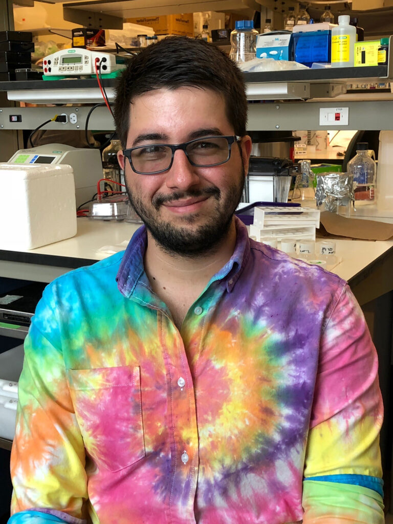
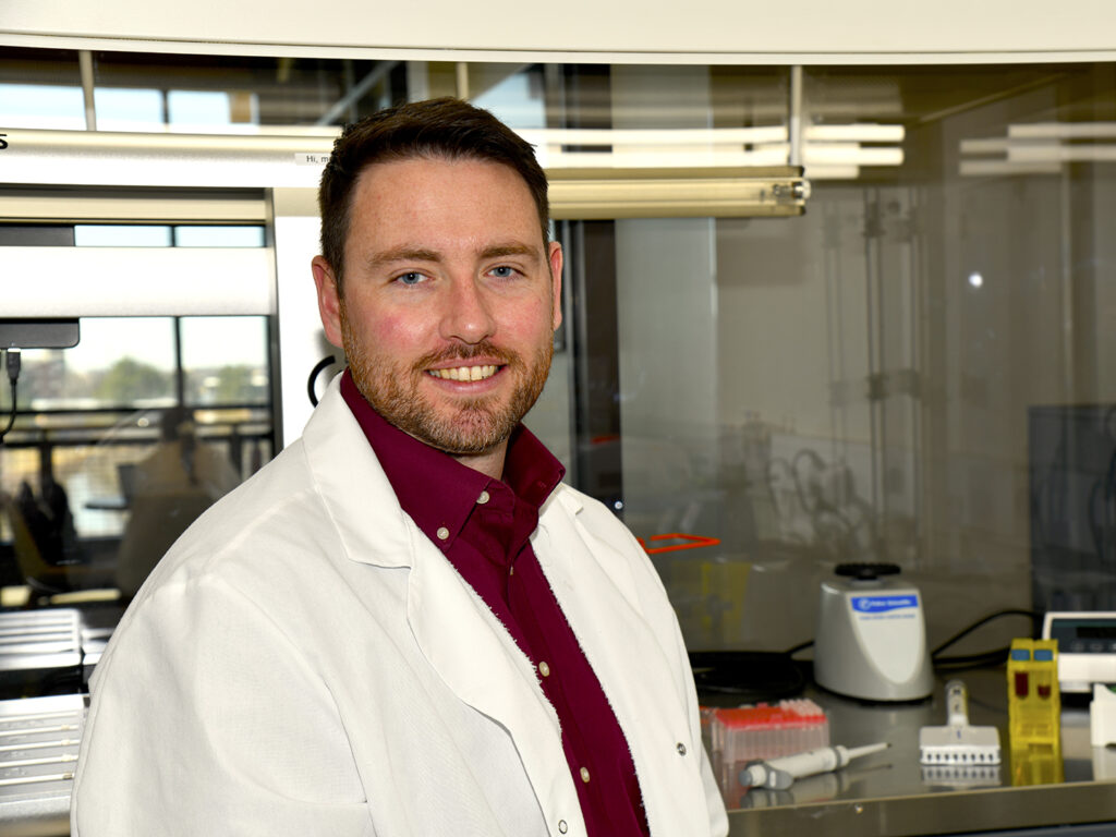
Pierre Ferrer, a Texas A&M School of Veterinary Medicine & Biomedical Sciences (VMBS) doctoral student, has been recognized with the American Society of Andrology’s 2023 Lonnie D. Russell Merit Award for his research on male fertility.
The Andrology Society of America in a top international scientific society supporting both medical and basic research on the male reproductive system. The Lonnie D. Russell Merit Award is given annually to a trainee member of the society who presented the best original laboratory or clinical research report.
During the society’s annual meeting in Boston, Ferrer presented his work on the protein mechanisms essential for spermatogenesis, or the production and development of mature spermatozoa.
He found that the actin-like 7A (ACTL7A) protein regulates several factors necessary for the formation of the acrosome, an organelle at the tip of a sperm that allows them to penetrate an egg.
ACTL7A is unique to the male reproductive system and extremely important; absence of or mutations in this gene causes infertility.
“Understanding the roles of proteins like this puts us a step closer to treating infertility and developing viable contraceptives for men,” Ferrer said. “I was surprised and very happy when they called my name (for the award); I have always looked up to Dr. Russell and his work in our field, so it is a personal honor to receive this award!”
Ferrer’s research was published in April in the journal “Molecular Human Reproduction.”
At the VMBS, Ferrer conducts research and trains in the lab of VMBS assistant professor Dr. Tracy Clement, whose work focuses on the genetics, epigenetics, molecular biology, and cellular biology involved in male fertility.
“It has really been a joy to see Pierre come into his own in research and fruitfully apply research methods to investigate the mechanisms of actin-related proteins in male fertility,” Clement said. “Recognition of Pierre’s work through this merit award speaks to the quality, rigor, and impact of the work he is leading in the lab.
“Pierre has a sharp memory and is a naturally curious and creative problem solver,” she said. “I am very proud of his accomplishments and can’t wait to see all the great work he contributes throughout his career.”
###
For more information about the Texas A&M College of Veterinary Medicine & Biomedical Sciences, please visit our website at vetmed.tamu.edu or join us on Facebook, Instagram, and X.
Contact Information: Jennifer Gauntt, Director of VMBS Communications, Texas A&M College of Veterinary Medicine & Biomedical Sciences, jgauntt@cvm.tamu.edu,979-862-4216
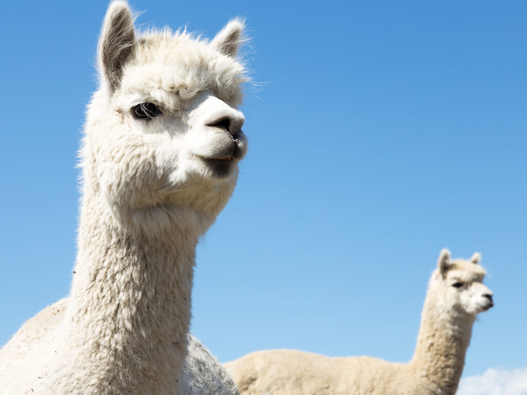
Cute faces, fluffy bodies, and calm attitudes are among the reasons alpacas have gained popularity as fun and unique pets.
“Many people choose to have alpacas as an alternative livestock animal,” explained Dr. Dusty Nagy, a clinical associate professor at the Texas A&M School of Veterinary Medicine & Biomedical Sciences. “They are relatively small and respect fences, which makes them easier to contain than many of our more traditional livestock species.”
Before beginning the alpaca ownership journey, however, owners should ensure they will be successful in caring for their alpacas by securing veterinary care and enlisting their veterinarian’s help in identifying healthy alpacas to buy.
Securing Veterinary Care
Veterinary care is essential to maintaining a pet’s health and resolving any emergencies that may arise. By finding a veterinarian before buying an alpaca, owners can guarantee they will be able to meet their alpaca’s health needs.
“Securing care for some species can be extremely difficult; not every large animal veterinarian will see alpacas, which is why owners should have a veterinarian before purchasing one,” Nagy said. “Having a veterinarian can be beneficial during the purchase as well, as they can provide appropriate confirmation that an alpaca is free from disease.”
Because gastrointestinal parasites are common in alpacas and can be deadly, Nagy recommends having your veterinarian perform a fecal parasite exam to be sure that there is a low amount of parasite eggs.
“Unless animals are housed in a dry lot, it is unrealistic to expect them to have no eggs in the feces on a consistent basis, so the test ensures that the level of parasite eggs is low enough for animals to remain healthy,” Nagy explained.
Benefits Of Owning Two Adult Alpacas
In addition to buying physically healthy alpacas, potential owners should avoid purchasing only one alpaca because alpacas are social herd animals by nature and tend to remain emotionally and physically healthy as part of a herd.
“It is not a guarantee that a single alpaca will bond with other animals that they are housed with, and not having a herd dynamic may leave them disengaged from the other alpacas,” Nagy said. “A herd also provides nurturing between alpacas and safety from predators. The lack of both can be stressful, and chronic stress can lead to illness.”
To foster a herd dynamic, Nagy encourages owners to purchase at least two alpacas that know and seem to like each other.
“The easiest way to find two animals that like each other is to get them at the same time from one breeder,” Nagy said. “Bonded animals typically stick close to one another within a herd setting. Most people who are observant of their herds know who these animals are and can guide people before a purchase.”
Potential owners may be interested in raising two young alpacas, specifically, but Nagy points out that raising young alpacas without an adult herd can result in emotionally unhealthy adults.
“Juveniles can develop significant behavioral abnormalities, such as aggression, when raised without an adult herd,” Nagy said. “For this reason, I recommend those new to alpaca ownership purchase only adult alpacas instead of young ones that need to be raised.”
Ultimately, prospective alpaca owners should not let an alpaca’s inviting appearance detract from their needs. Prioritizing their veterinary care and providing a herd is still essential to keeping alpacas healthy, allowing you to enjoy their wonderful companionship.
Pet Talk is a service of the College of Veterinary Medicine & Biomedical Sciences, Texas A&M University. Stories can be viewed on the web at vetmed.tamu.edu/news/pet-talk. Suggestions for future topics may be directed to vmbs-editor@tamu.edu.
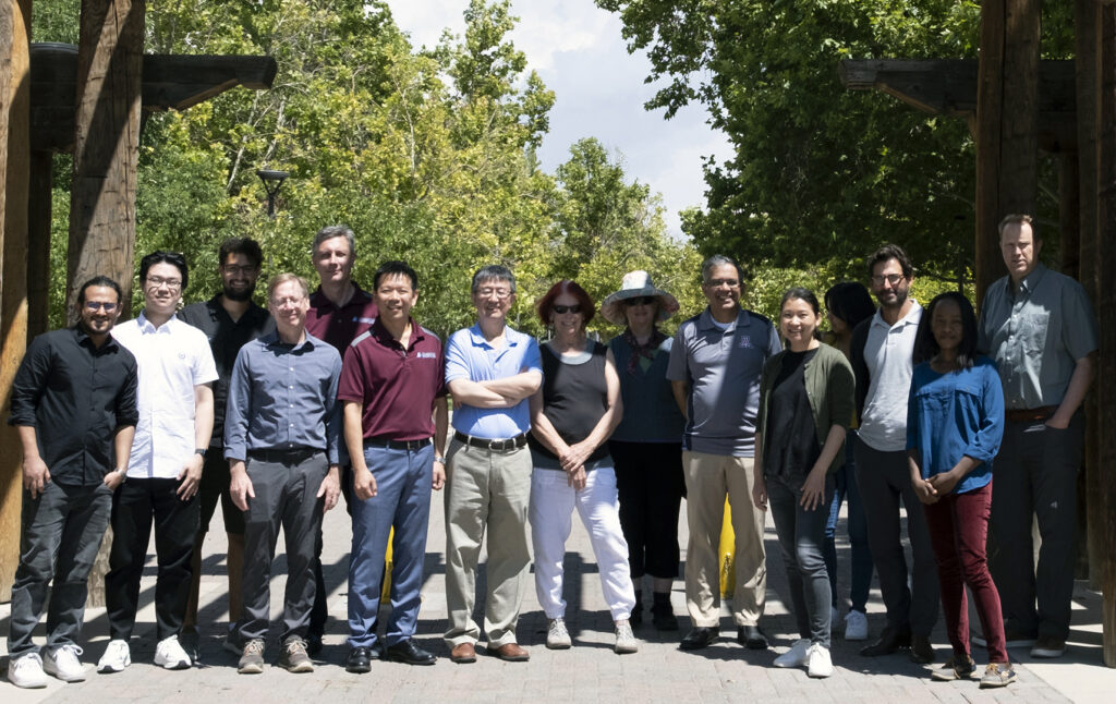
Summit attendees from the Texas A&M University, University of New Mexico, and University of Arizona Superfund Research Centers Photo by Theodros Woldeyohannes, University of New Mexico
Three Superfund Research Centers funded by the National Institute of Environmental Health Sciences (NIEHS) recently came together to share their accomplishments and challenges in fulfilling the data management and analysis mandate of the NIEHS Superfund Program.
Scientists from Texas A&M University, the University of New Mexico, and the University of Arizona met in Albuquerque for a day-long summit to kick off a regional collaboration of Data Management and Analysis Cores (DMACs). This partnership will identify opportunities to enhance data science, research translation, training, and community engagement, while simultaneously creating efficiencies in how the centers accomplish their goals in a challenging funding environment.
The Texas A&M University Superfund Center DMAC has implemented a rigorous system for quality assurance of research resources, data, and results. In addition, the core offers a number of unique high-dimensional analysis approaches that allow for exploration of the linkages between environmental exposure and adverse health effects data on both individual chemicals and mixtures.
“Our center’s focus on disaster research response creates a number of challenges with data management and analysis,” said Dr. Ivan Rusyn, center director and University Professor at the Texas A&M School of Veterinary Medicine & Biomedical Sciences. “The analytical solutions, focus on rigorous quality assurance, and training are not only contributing to the success of our own projects, but also benefiting the larger environmental science community and our community and government partners.”
The University of New Mexico Superfund Center DMAC, which hosted the summit, has unique expertise in managing and analyzing the data from a number of projects aimed at addressing environmental concerns of Native American communities. Strengths include enabling data-intensive projects with highly sensitive and privacy-protected environmental and population cohort data, as well as using geospatial approaches to report-back the results to the partner communities and the regulatory agencies to facilitate reduction in the burden of environmental pollution and ultimately improve the health of humans, animals, and the environment.
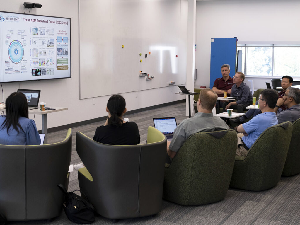
Summit attendees learning about the Texas A&M Superfund Research Center. Photo by Theodros Woldeyohannes, University of New Mexico
“The complexity of risks faced by our Native community partners has led us to develop novel approaches for analysis of mixtures that include psychosocial, dietary, and physiological risks that combine with contaminant mixture exposures,” said Dr. Johnnye Lewis, center director and research professor in the UNM College of Pharmacy. “We work within potentially conflicting needs of data security and privacy conferred by tribal data sovereignty juxtaposed with NIH needs of FAIR datasets that allow for combined analyses of data collected from many investigators. Models and methods developed to meet those needs can apply to many other communities where environmental injustices and disparities combine to create similar needs.”
The University of Arizona (UA) Superfund Center DMAC is a national leader in creating open science tools for “big data.” The UA houses CyVerse, an open data platform with more than a hundred thousand users across the United States and beyond that provides scientists with data storage, bioinformatics tools, data visualization, interactive analyses, cloud services, APIs, and more.
“Our Center leverages CyVerse to manage and analyze data from research projects that focus on exposures, health impacts, and mitigation of hazardous dusts in drylands,” said Dr. Xinxin Ding, center director, professor, and department head at the UA College of Pharmacy. “CyVerse supports the management and integration of data assets across our center, including establishing, coordinating, and monitoring processes for data management and analysis and identifying opportunities for integrating project/core-generated data with other existing datasets. Our DMAC team, including Nirav Merchant, CyVerse principle investigator and director of the UA Data Science Institute, is eager to introduce CyVerse to our regional partners and the rest of the Superfund Program.”
While each center has unique strengths in data management and analysis, the summit identified a number of areas where the centers can work together to leverage their strengths and promote rigorous, open, and collaborative environmental health science.
“Many of our graduate students and postdoctoral fellows could benefit from the combined expertise and tools the three programs have to offer,” said Dr. Matthew Campen at the University of New Mexico. “The diversity of research focus and applied data science approaches can create a more complete experience for our trainees.”
The centers agreed to create a Southwest Superfund Consortium in Data Science, a mutually beneficial partnership where each center’s DMAC agreed to establish communities of practice in data management/data science that deliver cross-center training, provide expert consultations, and share approaches and tools. Short- to medium-term priorities for the consortium will be in the areas of data privacy and security; quality assurance; geospatial data visualization and modeling; expanding the utilization of CyVerse in the environmental science and engineering projects; and training in open data science tools.
While ambitious, the consortium anticipates that these activities will not only create cross-center collaborations and efficiencies, but also ultimately result in enhanced solutions to environmental health issues that are plaguing a number of Native and environmental justice communities across the Southwestern U.S.
“While there are of course differences across centers, such as the more rural focus of UA and UNM compared to the more urban focus of Texas A&M, we believe the cross-fertilization of data science approaches across our centers will create synergies that ‘lift all boats’ in addressing the critical environmental health issues facing these communities,” said Dr. Weihsueh Chiu, the Texas A&M center’s deputy director.
###
For more information about the Texas A&M College of Veterinary Medicine & Biomedical Sciences, please visit our website at vetmed.tamu.edu or join us on Facebook, Instagram, and X.
Contact Information: Jennifer Gauntt, Director of VMBS Communications, Texas A&M College of Veterinary Medicine & Biomedical Sciences, jgauntt@cvm.tamu.edu,979-862-4216
Five trainees from the Texas A&M Superfund Research Center presented research at a webinar hosted by the Superfund Research Program’s (SRP) Student, Postdoc, and Alumni Network (SPAN).
The students participated in the Three-Minute Flash Talks portion of the webinar, which supplements graduate and advanced training by allowing trainees to present research to their peers while also developing presentation skills similar to those used at scientific conferences.
The School of Veterinary Medicine & Biomedical Sciences (VMBS) students who participated in the webinar included En-Hsuan Lu, Lucie Ford, and Hsing-Chieh Lin.
While some presented their contributions on the Texas A&M Superfund Center’s research projects, others presented their efforts related to the center’s support cores, with topics ranging from community engagement to data management.
Lucie Ford
The Superfund Center at Texas A&M is one of 25 centers at universities across the country funded by the National Institute of Environmental Health Sciences, a branch of the National Institutes of Health (NIH). The Texas A&M Superfund Center is unique from other centers in that its research doesn’t focus on just one class of chemicals but rather studies the whole picture of chemical effects during disasters.
This was evident in the topics the VMBS students presented at the webinar.
Lu and Lin are both second-year toxicology doctoral students who work on the Superfund Center’s Risk & Geospatial Sciences Core (RGSC) under Dr. Weihsueh Chiu, professor in the VMBS’ Department of Physiology & Pharmacology (VTPP) and deputy director of the Superfund Center. The RGSC is tasked with providing data and services that characterize human health risks and mapping geographic distributions of environmental mixtures created during disasters.
Hsing-Chieh Lin
Lu and Lin’s presentations advocated using human and computer-based models to gather toxicity data necessary for predicting human health risks and outcomes during disasters. Lin shared that the new models will help fill in the gaps in available toxicity data.
Ford is a third-year doctoral student in the toxicology program who works on the Superfund Center’s Project 4, led by Dr. Ivan Rusyn, VTPP professor and Superfund Center director. Project 4 responds to the Superfund mandates for advanced techniques for detecting, assessing, and evaluating hazardous substances, as well as developing methods to assess health risks.
Also representing the Texas A&M Superfund Center was Dr. Eva Vittuci, a postdoctoral research associate on Project 2, led by Dr. Natalie Johnson, vice chair of the Interdisciplinary Program in Toxicology and an Associate Professor in School of Public Health. This project focuses on measuring air pollution using a state-of-the-art scientific vehicle on site during disasters.
Eva Vittuci
Vittuci’s presentation discussed the team’s efforts in developing new tools that can rapidly characterize volatile organic compound (VOC) mixtures. VOCs can vaporize into air, making them easy to inhale. People who are exposed to VOCs can suffer a variety of health effects, including respiratory issues and organ damage, depending on the compound, exposure level, and timeliness of medical care.
Greg Kudzin is a doctoral student in chemistry at the University of North Carolina at Chapel Hill who works on the Superfund Center’s Project 1, led by Dr. Erin Baker, an associate professor of chemistry at UNC Chapel Hill. Project 1 aims to develop novel analytical and computational methods for detecting and assessing hazardous substances.
Kudzin’s presentation was on methods of detecting the presence of per- and polyfluoroalkyl substances (PFAS). PFAS are substances used in a variety of household products since the 1940s that can contaminate the water supply in urban areas. They are often called “forever chemicals” because they don’t naturally decay. Once they enter human bodies or the environment, they’re often permanent.
Greg Kudzin
Not only did the presentations allow the Texas A&M Superfund Center trainees to practice their presentation skills but it also allowed them the opportunity to share their hard work with peers and experts from the toxicology community. Given the unpredictable nature of disasters, it’s vital that researchers build collaboration networks that can work together to solve global problems.
For more information about the Texas A&M College of Veterinary Medicine & Biomedical Sciences, please visit our website at vetmed.tamu.edu or join us on Facebook, Instagram, and X.
Contact Information: Jennifer Gauntt, Director of VMBS Communications, Texas A&M College of Veterinary Medicine & Biomedical Sciences, jgauntt@cvm.tamu.edu,979-862-4216
Recently, Texas Gov. Greg Abbott announced a wildfire disaster declaration for about 75% of the state’s counties and the National Oceanic and Atmospheric Administration also released an updated hurricane season outlook that includes a 70% chance of 14-21 named storms before the season ends in November.
With these potential weather threats in mind, now is a good time to consider what you might do with your pets if there was an emergency.
Having a plan in place is crucial for the furry members of your family. The most important way for owners to prepare for a disaster evacuation is to plan ahead and pack a go-kit, an easy-to-grab, waterproof bag or container with basic survival items and supplies.
Guidance On Planning Ahead
The first step in planning ahead is to know where you are going in the event of a possible evacuation by compiling a list of hotels, boarding facilities, and shelters that allow pets.
“This helps you know where you can go, depending on the types of animals you have and how many, and can give you a leg up on calling them quickly when you know you will be evacuating and need a reservation,” said Dr. Deb Zoran, a professor at the Texas A&M School of Veterinary Medicine & Biomedical Sciences. “Before you need them, be sure you have information on the hotel, shelter, or boarding facilities you may use when a disaster is headed your way, check that they have a good reputation for their animal care, and make sure the facilities are completely out of harm’s way.”
While gathering information, owners can also confirm if the facilities require specific vaccinations.
“The only vaccine that is required by law in most places is rabies, yet even that vaccine is not always required for entry into an emergency animal shelter,” Zoran said. “A majority of animals brought to a shelter after being rescued from a flood, fire, or tornado come without their owners – or their owners come with them but without documentation. Emergency shelters have to operate on this premise.”
To ensure there is no doubt about a pet’s vaccination status, owners should keep records of vaccinations that evacuation shelters may require evidence of, including:
Rabies
DA2PP, a single vaccination for distemper, adenovirus type 2, parvovirus, and parainfluenza
kennel cough for dogs
FVRCP, or feline viral rhinotracheitis, calicivirus, and panleukopenia
“Most shelters allow non-vaccinated animals into the shelter but separate the animals with vaccination papers from those without,” Zoran continued. “Ultimately, you need to consult with your vet about appropriate vaccines for your dog or cat.”
Assembling A Go-Kit
Owners should ask their veterinarian annually for copies of vaccine records and medication details to print and include with their go-kit.
“If your pet needs medications – heartworm, flea prevention, or specific medication for health conditions such as thyroid or heart medicine, antibiotics, etc. – you need a copy of the medical records showing that your pet requires this medication should you not have enough or the medication is lost and has to be refilled,” Zoran explained. “Pet medications cannot be given to you without documentation or a new vet exam, so having records available is important.”
A go-kit should also include first aid supplies; cleaning supplies, including pet waste bags and sanitizing wipes; and feline supplies, if applicable, such as a litter box, scooper, and litter.
Zoran suggests owners pack additional items that can help keep pets safe and comfortable while traveling and throughout their stay in an unfamiliar place.
“Disasters are a stressful time for pets, so bringing things that smell familiar, special treats, normal food, and toys is very important,” Zoran said. “Cats, in particular, not only need the kennel that they travel in, but if they are going to be away from home for days or weeks, they will need a kennel at least 2 feet by 3 feet or larger to live in; they need room to sleep, a box to hide in, and a place for the litter tray.”
Normal foods that pets are accustomed to contribute to keeping pets healthy during difficult situations.
“Familiar foods and water sources will help prevent gastrointestinal upset, as sudden food changes in the shelter environment can lead to loss of appetite, vomiting, or diarrhea, adding to the stress of the moment,” Zoran said. “Cats will be less willing to eat in a stressful or busy environment if they don’t have their own food and if they don’t have a quiet place or time to eat.”
Disaster evacuations can create stressful environments for people and pets alike, but adding identification tools and transportation supplies into a go-kit can help prevent pets’ needs from being overlooked in the chaos.
“Both dogs and cats need to have microchips, collars, or harnesses for identity protection and escape prevention,” Zoran explained. “Cats should not be removed from kennels without a harness on, as fear-inducing experiences can cause them to run and hide. Dogs, even the most highly trained ones, will also flee, so careful use of collars and leashes is essential, both for their protection and for preventing interactions with other animals at the emergency shelter.”
When disaster strikes, household pets are among the most vulnerable, as they depend on us for care. By never leaving them behind, staying informed, and, most importantly, preparing ahead for an emergency evacuation, you can keep your furry friend protected, safe, and healthy throughout any disaster.
Pet Talk is a service of the School of Veterinary Medicine & Biomedical Sciences, Texas A&M University. Stories can be viewed on the web at vetmed.tamu.edu/news/pet-talk. Suggestions for future topics may be directed to vmbs-editor@tamu.edu.
All Welcome Week events are MaroonBase eligible. Students who download the app, sign up, and check into the events can earn points to win prizes. The VMBS group code is 150255229276.
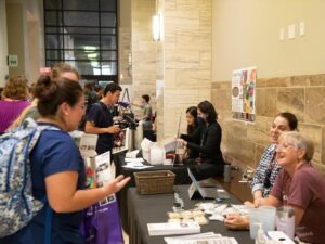
Resources Fair
Monday, Aug. 21 | 3-6 p.m. VENI Grasshopper Room Hallway
The annual resources fair is designed to introduce new and existing campus members to the variety of well-being and academic support resources available at the VMBS and across Texas A&M University. This event is open to all of West Campus.
Bingo! Mixer
Tuesday, Aug. 22 | Noon to 1 p.m. VENI Third Floor Commons
It’s not your grandparents’ Bingo! Make new connections and learn fun facts about your friends at the VMBS’ annual Bingo! Mixer. Snacks will be provided. This event is sponsored by VOICE (Veterinarians for One Inclusive Community for Empowerment).
Safety Training with Texas A&M UPD
Tuesday, Aug. 22 | 5-7 p.m. VIDI 103
Learn valuable self defense techniques from the Texas A&M University Police Department (UPD) in this interactive training class. Bring comfortable clothes and shoes and be prepared to sweat!
Are you a first-generation college student or interested in learning more about the unique challenges these individuals face? Attend the panel to make connections, hear stories from first-gen students and faculty, and enjoy a sweet treat.
Why is mentorship such an important part of higher education and veterinary medicine, in particular? Learn from a panel of faculty, staff, and students and find out what mentorship can do for you. Dessert will be provided.
Enjoy a fun Friday dessert while learning wellness tips and tricks Mike Hawkins, the Veterinary Medical Teaching Hospital’s licensed professional counselor.
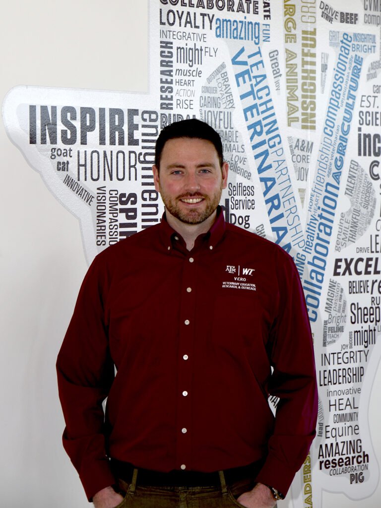
Rather than being a veterinarian like many of his VERO colleagues, Pinnell is an environmental microbiologist who has spent his education and career studying much smaller organisms — the microscopic bacteria and other living creatures that can be found everywhere from soil and ocean water to our own digestive tracts.
“What I like about the microbial ecology field is that you can take the tools, skills and concepts and really apply them to anything,” Pinnell said. “It’s all the same kind of stuff; it’s just wrapped in a different context. I’ve jumped all over the place as far as environments and hosts go.”
Now, as a research assistant professor at the Texas A&M School of Veterinary Medicine & Biomedical Sciences (VMBS), he’s using his diverse skills and experiences to help tackle some of the most important issues in the cattle production industry.
From The Ground Up
Pinnell grew up in a small town in central Ontario, Canada, before moving south to study at the University of Waterloo.
He had his first experience with research while completing a bachelor’s degree in biology — working with aquatic ecologists to study zooplankton and invasive zebra mussels in Ontario.
Then, during a field course in the Bahamas between his third and fourth year, Pinnell stumbled into the field of microbial ecology.
“One of the faculty members on the trip, Josh Neufeld, did microbial ecology work and asked a buddy and me if we wanted to work with him as master’s students,” he recalled. “My master’s work was looking at microbial communities in tundra permafrost as part of a larger project that was biofuel-focused.”
After completing his master’s degree in 2011, Pinnell decided to take a break from academia and began working as a laboratory technician at The Hospital for Sick Children (SickKids) in Toronto.
“The best thing about VERO is the team. It has a very family-style feeling.”
Dr. Lee Pinnell
While studying Inflammatory Bowel Disease (IBD) and Crohn’s disease in one of the hospital’s gastrointestinal labs, Pinnell had his first experience investigating the gut microbiome, the community of microorganisms inside the digestive tract that convert food to energy and play a big role in the body’s overall physical and mental health.
After a couple years in the lab, Pinnell began to miss the opportunities for creativity that academia provides and started searching for a Ph.D. program in which he could expand his knowledge in computational biology and marine microbial ecology.
That search led him to Texas, where he began a doctorate in marine biology at Texas A&M University-Corpus Christi.
During Pinnell’s first visit to Corpus Christi, he was exposed to the pollution that plagues the Gulf Coast of Texas.
“It had rained really hard for a couple days, and in Corpus Christi, the water just flows down the streets and comes out of the storm drains into the bay,” he said. “Jeffrey Turner, my faculty supervisor, and I were driving along and saw that the shoreline and waters of Corpus Christi Bay were just littered with plastic. Jeff and I thought it’d be interesting to do a project on plastic pollution and how it’s impacting microbial communities in the area.”
In the Laguna Madre estuary, the body of water between Padre Island and the mainland where fresh and saltwater mix, the team exposed local microbial communities to plastic, biodegradable plastic and ceramic to study how the microbes colonized on and reacted to each material.
They found that the microbes in the biofilms on the biodegradable plastic tended to contain more sulfate reduction and antimicrobial-resistant genes than those on the typical plastic.
“So, while bioplastic is better from an environmental loading perspective because it’s being degraded when plastic is not, it’s still changing environmental processes,” Pinnell said. “It begs the questions whether bioplastic should also be considered a pollutant, since it’s changing baseline values in the environment.”
After spending about a year at the Shedd Aquarium in Chicago studying how probiotics impact beluga whales’ microbiomes, Pinnell found a postdoctoral research opportunity with Dr. Paul Morley, the director of food animal research at VERO.
Joining The VERO Family
Pinnell never expected to end up joining a program focused on production animal health.
“I didn’t even know animal science was a thing before 2021. We didn’t have those programs at the schools I went to,” he said. “I don’t think I’d ever been to a dairy, and I’d definitely never been to a feedlot.”
The team-oriented research environment that Morley cultivates is what initially attracted Pinnell to the VERO program, but it didn’t take long before he also started to enjoy working in a field that few other environmental microbiologists had explored.
“There weren’t as many people taking microbial ecology concepts and applying them to veterinary science,” he said. “It just felt like there was a window where we could do something unique, which was really cool to me.”
He also realized that what he thought was a disadvantage — having little background knowledge on cattle health and the production industry — turned out to be an advantage.
“Because I was a traditional environmental microbiologist, I had no preconceived notions of what’s supposed to be the cause of Bovine Respiratory Disease (BRD) or liver abscesses in cattle,” he said. “At VERO, everybody has slightly different perspectives, which is part of the reason we work very successfully — we don’t see things exactly the same way.”
His postdoctoral research focused largely on cattle liver abscesses, a common and serious issue in feedlots, and the role that the gut microbiome plays in abscess formation. Since then, he has expanded his research to include the microbial ecology of BRD and antimicrobial resistance.
When a faculty position became available in February 2023, Pinnell jumped at the chance to find a more permanent position at VERO.
“The best thing about VERO is the team. It has a very family-style feeling,” he said. “I’m always telling younger grad students that when they’re looking for labs, who you’re going to work with and those relationships are more important than the actual project.”
In Pinnell’s case, however, he happened to find a place that offers both a great team and a great research focus.
“The applicability of the research — how closely we work with the people who can actually implement the findings — is great,” he said. “That’s what’s really cool about Canyon, you can easily go and talk to someone who’s dealing with these problems every day.”
Looking Forward And Setting Goals
Two of Pinnell’s priorities for his work at VERO include continuing to grow his own knowledge and beginning to train and mentor graduate students and postdoctoral researchers.
“I’ve been so lucky throughout my career in that I’ve had amazing supervisors, so I really want to do a good job mentoring my students,” he said. “It’s important to me that they have a good experience, both with the research and just in general. Because if you don’t like something, you’re probably not going to continue to do it for a career.”
He also plans to continue conducting meaningful research and implementing the outcomes of that research however possible.
“VERO does a really good job of communicating with non-scientists, because there are a lot of ties between VERO researchers and Texas Panhandle producers,” Pinnell said. “I want to make sure that people are aware of what we’re doing and that they have access to the findings.”
###
For more information about the Texas A&M College of Veterinary Medicine & Biomedical Sciences, please visit our website at vetmed.tamu.edu or join us on Facebook, Instagram, and X.
Contact Information: Jennifer Gauntt, Director of VMBS Communications, Texas A&M College of Veterinary Medicine & Biomedical Sciences, jgauntt@cvm.tamu.edu,979-862-4216
Routine vaccinations are an essential part of your pet’s veterinary care, and veterinarians typically guide owners on how often your pet needs which shots. In the case of rabies, however, owners may be asked by their veterinarian if they prefer a vaccine that lasts one year or three years.
“The differences between one-year and three-year vaccines depend on the specific vaccines,” said Dr. Lori Teller, a clinical associate professor at the Texas A&M School of Veterinary Medicine & Biomedical Sciences. “Many of the adjuvanted vaccines — or vaccines with an added chemical to boost immune response and ensure protection — are similar, with the only difference being the label. The biggest factor that owners should consider is what the law is in their jurisdiction.”
Local And State Laws On Rabies Vaccines
The beginning of the rabies vaccination schedule is the same for cats and dogs, which is important for owners to follow since rabies is a viral disease that spreads through bites or saliva. This infectious disease will affect the central nervous system and cause symptoms such as excessive drooling, aggression, seizures, and death.
There is no effective treatment for rabies, and because rabies can spread from animals to humans, vaccinations are necessary to protect both pets and their owners.
“Rabies is considered a core vaccine, meaning that all dogs and cats should be vaccinated for protection against this fatal disease,” Teller said. “A puppy or kitten should receive their first rabies vaccination between 12-16 weeks of age and then will require a booster vaccination 12 months later. Any subsequent vaccinations will be determined by what local or state laws mandate.”
In fact, local and state laws can differ depending on how severe the risk of rabies is in an area, making it crucial for owners to discuss local vaccination requirements with their veterinarian. These laws should at least outline how often a pet should be vaccinated and with which form of the rabies vaccine.
“Laws regarding rabies vaccination vary from state to state and from jurisdiction to jurisdiction,” Teller said. “Texas requires dogs and cats to be vaccinated against rabies, but some states do not require it while others require only dogs to be vaccinated. Some municipalities require pets to be vaccinated annually, others require biennial rabies vaccinations, and then many require triennial vaccinations. There are even some that require a pet to be vaccinated annually with the three-year vaccine.”
If a pet is found to be unvaccinated for rabies or has bitten someone, owners can be fined or their pet may be quarantined away from home until it is confirmed they are not infected. Pets are also typically required to be vaccinated if they are around other animals, such as in a boarding facility or grooming salon.
Rabies Vaccine Requirements Depending On Pet
As long as local and state guidelines on rabies vaccines are met, owners may be given the option to choose between the one- or three-year forms of the vaccine. These options are dependent on the type of pet, according to Teller.
“If the law says that a pet can receive a three-year rabies vaccination every three years, then that would be appropriate for most dogs,” Teller explained. “The caveat to this is for cats because many veterinarians use the feline-specific, non-adjuvanted vaccine, and they prefer the one-year version for cats because it is less expensive than the three-year version and is less likely to cause short-term side effects, such as low-grade fever or tenderness at the vaccination site.”
Working with your veterinarian to determine which rabies vaccine is right for your furry friend will keep them safe and healthy while also preventing the disease from spreading in your local community.
Pet Talk is a service of the College of Veterinary Medicine & Biomedical Sciences, Texas A&M University. Stories can be viewed on the web at vetmed.tamu.edu/news/pet-talk. Suggestions for future topics may be directed to vmbs-editor@tamu.edu.
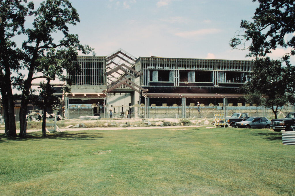
Current Large Animal Teaching Hospital facilities under construction
In December 1888, a small, wood-frame barn was built at Texas A&M University to serve as a veterinary hospital. Ever since that original, 720-square-foot barn served the university’s first veterinary patients, the campus has been home to continuously improving veterinary facilities.
When the School of Veterinary Medicine & Biomedical Sciences (VMBS) was officially established in 1916, it took on the responsibility of running the Veterinary Medical Teaching Hospital facilities. Today, the VMBS is home to the state’s only veterinary teaching hospital consisting of the Large Animal Teaching Hospital (LATH) and the Small Animal Teaching Hospital (SATH).
The current LATH facility, which began serving patients in August 1993, plays an important role in treating horses, cattle, and all other hoofstock with its state-of-the-art design specially built to meet the unique needs of modern large animal veterinary medicine.
This year, the facility celebrates 30 years of housing the exceptional people who provide cutting-edge medicine to large animals throughout Texas and across the United States, as well as veterinary innovation, groundbreaking research, and outstanding education to future Aggie veterinarians.
“These walls see the students, staff, and faculty who are committed to doing the best we can for every patient that walks in the door and to providing compassionate care at the expert level expected for our patients,” Schleining explained.
Dr. Stacy Eckman, associate dean for hospital operations, echoed Schleining’s enthusiasm for the bright and talented minds who’ve worked within the LATH’s walls.
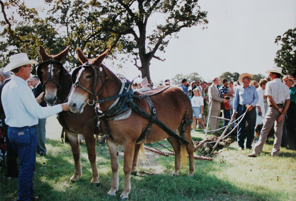
Groundbreaking of the Large Animal Teaching Hospital facilities
“Our faculty foster a welcoming environment for all students, and I think that’s a really important part of what happens here,” Eckman said “They are willing to take on the toughest of the tough cases. They’re willing to be innovative. They’re willing to drive the next generation of veterinarians to push themselves to further expand the boundaries of what we can offer here.”
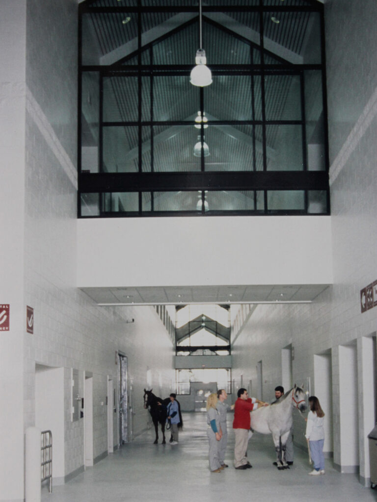
It’s hard to walk through the LATH without feeling a sense of awe, both because of the brilliant veterinarians who work there and because of the layout and design of the building. The building exudes a rare combination of spaciousness, ease, and warmth.
“I think the proximity with which we all interact within the space without being cramped and feeling like we don’t have to compete for resources is one of the special parts, but it is also the pride of being part of something as special as Texas A&M within the walls of this well-built hospital,” Schleining said. “That pride really connects people to want to do good things with each other here.”
Veterinary Innovation
The spacious, high-ceilinged corridor makes caring for large patients safer for the animals and those providing their care.
The LATH serves as a reminder of just how far veterinary medicine has come since the university began serving patients out of a small wooden barn 135 years ago.
“Our operating rooms are state-of-the-art, and our flooring system is specially designed and padded to help protect our hoofed patients coming out of surgery,” Eckman said. “Some of our rooms have extra room so that a partner horse can be by a patient’s side to provide support. These are just a few of the special needs our LATH so thoughtfully includes.
“The LATH also includes several design elements inspired by Temple Grandin, an internationally recognized animal behaviorist, to help minimize a patient’s feelings of anxiety or fear as they enter a new or unknown space,” she said.
The facility’s innovative design also encourages collaboration, Schleining shared.
“Even though we have a large space, we fill it with dedicated people who are always conversing and collaborating within the LATH halls, making magic happen,” Schleining said. “We are really blessed to have the space to be able to provide the most advanced compassionate care for large animals and to have a facility designed in a way that encourages people to gather and work through challenges together.
“A recent example of this is Dante, a longhorn steer who came to the Food Animal and Surgery Service in the LATH from the Austin Zoo with a fractured skull,” she continued. “His case started in radiology and ended with an innovative, first-of-its-kind surgery with input from food animal surgeons and medical internists, radiology, orthopedics, anesthesiology, and our equine farrier who was able to fabricate materials for us.”
This kind of collaboration is a daily occurrence at the LATH, which is why clients have traveled from 37 of the 50 United States seeking veterinary care at the facility.
Groundbreaking Research
With innovation comes research, and the LATH helps facilitate research aimed at improving large animal veterinary medicine.
“The world’s best hospitals — like the Mayo Clinic, Johns Hopkins, Mass General, etc. — are places where clinical research is actively advancing biomedical research to improve patient care,” said Cohen, who also serves as the VLCS’ associate department head for research and graduate studies. “The end goal of biomedical research is to improve the health of people and animals. This is ultimately manifested in applying new tests to better identify individuals with disease or at increased risk of disease, improved treatments for patients with disease, and preventatives like vaccines to stop disease from developing. The faculty and staff who work in the LATH conduct this work that enables us to provide cutting-edge care.”
The LATH facilities also add to an environment that promotes observational studies, or research in which patient outcomes are examined overtime.
“The records of our patients can be reviewed in observational studies to help us learn about which interventions work best for patient management, which findings predict better clinical outcomes, and which historical findings or tests best inform diagnosis,” he shared.
Cohen said the research that the LATH facilities help make possible also enhances the student experience and the overall quality of care provided.
“Our students can learn about the impact of translational research in advancing veterinary medicine,” he said. “The collective, team-based approach of the LATH enables us to provide the highest quality care for our patients, including the application of innovations from diagnostic tests, treatments, and preventatives developed at Texas A&M and beyond.”
Outstanding Education
Dr. Kevin Washburn and students examine a heifer’s eye.
In addition to encouraging innovation and research, the LATH’s architectural design also enhances learning and teaching efforts that take place there by providing more room for visual and hands-on learning.
“In the vast majority of the clinical rotations here, (fourth-year) students get a lot of hands-on experience and are honing skills that they can take forward with them after they graduate,” Schleining explained. “The intentionality of this building helps make that possible.”
Eckman’s was among one of the early Doctor of Veterinary Medicine classes to learn in the current LATH and said it provides a much better learning environment than previous hospital buildings.
“The teaching and learning experiences really benefit from the larger exam rooms,” she said. “The older facilities were much more cramped, which made it harder to see what you were supposed to be learning. That’s certainly not the case now. We have multiple options for safely viewing and learning. For example, in our recovery stalls, when a patient is coming out of anesthesia, we can have students safely watching on the catwalk above.”
A Building Worth Celebrating
The LATH has played an important role in advancing human and animal health by enhancing the veterinary educational experience over the last 30 years. Eckman said she’s excited to celebrate this important milestone.
“Buildings wear down over time, but from a space standpoint and our ability to teach and care for patients in that building, it really hasn’t aged. We still offer state-of-the-art care and continue to offer new and improved treatments in the LATH,” she said. “It’s worth celebrating because this building has withstood the test of time.
“As we look forward to the next 30 years in the LATH, we will continue to have a sense of adventure when taking on challenging cases and thinking outside of the box on how we can improve the life of a patient, whether they are a working animal or a beloved pet.”
###
For more information about the Texas A&M College of Veterinary Medicine & Biomedical Sciences, please visit our website at vetmed.tamu.edu or join us on Facebook, Instagram, and X.
Contact Information: Jennifer Gauntt, Director of VMBS Communications, Texas A&M College of Veterinary Medicine & Biomedical Sciences, jgauntt@cvm.tamu.edu,979-862-4216
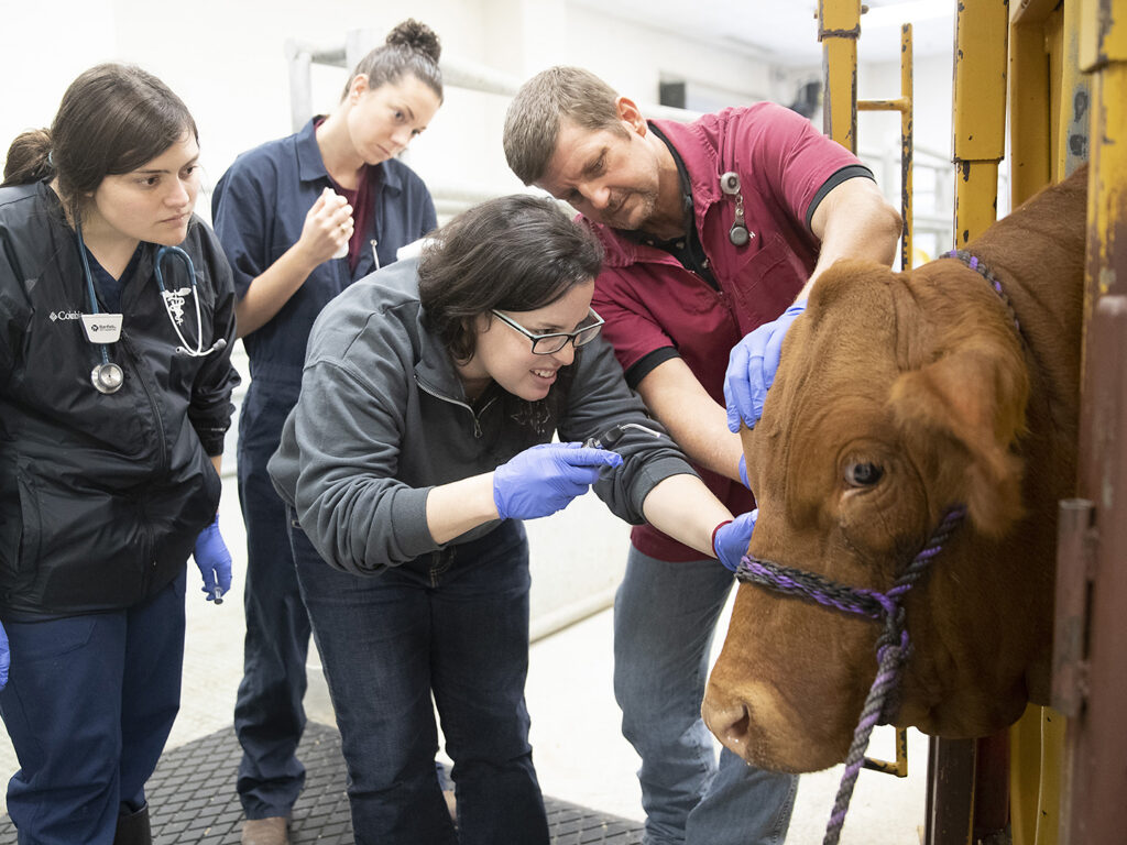
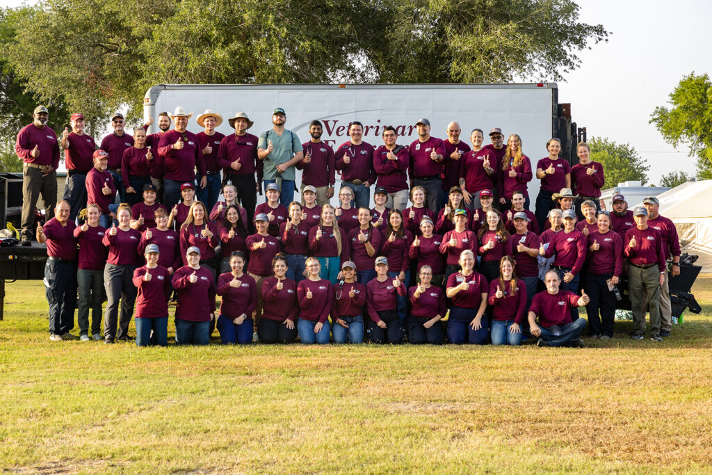
In the early hours of July 22, long before the sun began its ascent over the Brazos Valley, cars began turning into The Texas A&M University System RELLIS campus.
Early risers gathered to join the Texas A&M Veterinary Emergency Team (VET) on its journey to South Texas for Operation Border Health Preparedness (OBHP), an annual readiness exercise hosted by the Texas Department of State Health Services that provides emergency response teams the opportunity to test their readiness for the next major disaster while also providing annual medical and veterinary care to communities that would otherwise go without it.
The team worked throughout the weekend to establish a base of operations at Raymondville Early College High School with the Texas A&M AgriLife Extension Service Disaster Assessment and Recovery (DAR) team, which provides logistical and administrative support to the VET during deployments.
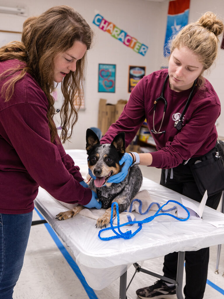
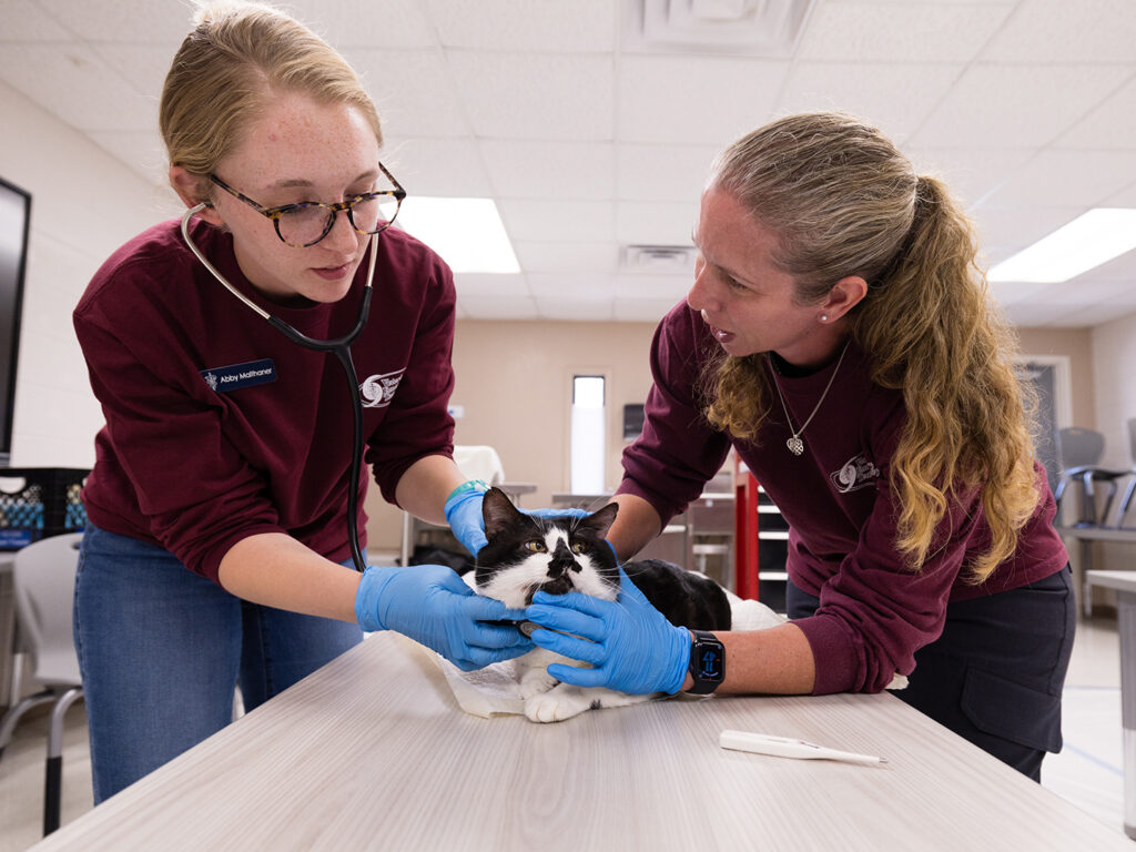
The group’s efforts in setting up tents, pet-proofing classrooms and practicing with the team’s medical records system were met Monday morning with enthusiastic Texans eager to receive veterinary care for their beloved cats and dogs.
By the end of day one, the VET had served 119 patients. By the end of the week, the team had completed a record 1,022 veterinary visits at its base of operations in Willacy County, which almost doubled the number of patients the team saw in 2022 and is significantly higher than their caseload in 2021, the team’s first year to participate.
“Willacy County is historically the poorest in Texas and the fifth poorest in the nation, with no veterinarian serving the community,” said Dr. Wesley Bissett, VET director. “But the people here love their animals as much as any other Texan, and their animals deserve care. We’re honored to have the opportunity to provide that care at OBHP at no cost thanks, in part, to the generosity of organizations like the Banfield Foundation.”
Sixteen College of Veterinary Medicine & Biomedical Sciences Doctor of Veterinary Medicine students joined the VET at OBHP in 2023, giving them hands-on experience in providing veterinary medicine in a disaster exercise. Daniel Martinez, a fourth-year veterinary student originally from the Rio Grande Valley, was among the students who participated in OBHP.
“The Rio Grande Valley is a poverty-stricken area of Texas where citizens live in multi-person households, with median household incomes that look like the price tag of a modest used car, about $40,000 in Willacy County,” Martinez said. “Regardless, clients were bringing in their pets to us, sacrificing their resources to make the trip to Raymondville Early College High School where the VET and DAR provided a service deemed essential to the community.”
Dr. Deb Zoran, a professor of small animal medicine and founding member of the VET, teaches the two-week VET rotation course to fourth-year students when she’s not deployed with the VET or providing medical care as the veterinarian to the Texas A&M Task Force 1 urban search and rescue dogs.
“The VET rotation is built like every other two-week rotation in the school,” Zoran said. “We split the rotation into one week of preparedness and one week of response. As it turns out, OBHP happens to fall on a response week, so students on the VET rotation during OBHP don’t just practice in a virtually simulated deployment; they get to participate in a full-scale disaster response exercise with hands-on participation in establishing a base of operations and providing basic veterinary care.”
The experience is invaluable.
Martinez said he saw the best of humanity in his mentors, fellow students and clients at OBHP.
“I built relationships with my classmates, teachers and other personnel that demonstrate the core values of Texas A&M University,” Martinez said. “The VET and DAR working together was a workshop in teamwork. The veterinary profession cannot exist without teamwork. What happened in Raymondville is just a snapshot of what can happen when caring individuals come together.
“This year’s Operation Border Health Preparedness added value to my professional veterinary medical education by underscoring the synergy of the veterinary profession and the demographic it serves,” he said.
One of the families that brought its pets to OBHP for basic veterinary care echoed Martinez’s admiration for the services provided by the VET and DAR. The Gonzalez family adopted two calico kittens at the beginning of the COVID-19 pandemic, naming them Snickers and KitKat.
“The initial thought was that they’d be animals who helped keep our kids sane during the early challenges of COVID, but they quickly became much more than that — they’re two additional members of the family,” said Roel Gonzalez, a math teacher at Raymondville Early College High School. “They really care about my kids, and my kids have so much affection for them. It’s nice to be able to bring them here for veterinary care so that we can take care of them the way they’ve been caring for us over the last few years.”
The VET is able to focus on caring for family members like Snickers and KitKat on deployments rather than having to expend the team’s energy on some of the more logistical elements of a deployment, thanks to help from the DAR team.
“We deploy in cooperation with the VET to help with logistical support,” said Rachel Bauer, the liaison to Texas A&M AgriLife Extension at the state operations center in Austin who also works with the Texas Division of Emergency Management. “Our AgriLife Extension Disaster Assessment and Recovery agents free up the VET members to focus on providing the best veterinary care possible. We do this by working to keep the generators running, greeting the public and guiding them on where to go once they’ve entered the VET’s base of operations, and providing any other logistical support they may need.”
Dr. Monty Dozier, the program director for the DAR team, said OBHP is a meaningful experience because it builds connections with people and organizations across the state while furthering the connection between DAR and the VET.
“Our partnership with the Texas A&M VET is one that we value highly, and OBHP is an opportunity for us to work together to make things better for Texas,” he said. “We get to practice our techniques for training and working with volunteers, which is important because during a disaster response, we take in volunteers and train them on how to contribute to our efforts.”
Bissett and Zoran said the DAR agents who deploy with the team make their operations smoother while adding an element of comradery.
They also pointed out that the VET would not be able to provide the care and services it offers at OBHP without generous donors. Bissett estimated that the care the team provided at no-cost to South Texans during OBHP would have equated to $400,000 in out-of-pocket expenses at a traditional veterinary clinic.
“OBHP is not possible without the support we receive from our donors; they make it possible to provide veterinary care to this special community,” Zoran said. “We are beyond thankful to our donors for seeing the importance of making veterinary care more accessible and the value in investing in the VET to make that possible at OBHP.”
###
For more information about the Texas A&M College of Veterinary Medicine & Biomedical Sciences, please visit our website at vetmed.tamu.edu or join us on Facebook, Instagram, and X.
Contact Information: Jennifer Gauntt, Director of VMBS Communications, Texas A&M College of Veterinary Medicine & Biomedical Sciences, jgauntt@cvm.tamu.edu,979-862-4216