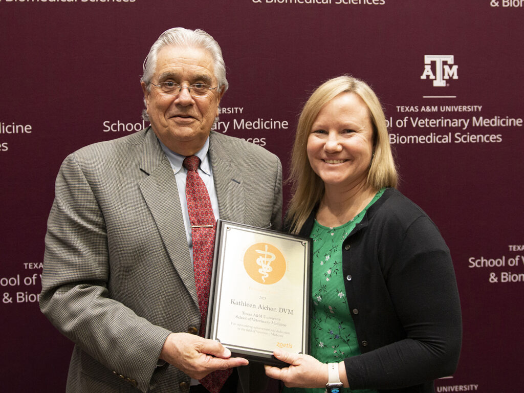
The Texas A&M School of Veterinary Medicine & Biomedical Sciences (VMBS) recognized 72 students, faculty, and staff at the 2023 Honors & Awards Ceremony on April 6.
“It is always an honor to recognize our colleagues who are leaders in the veterinary profession, committed to educational excellence, and provide dynamic leadership,” said Dr. John R. August, the Carl B. King Dean of Veterinary Medicine at Texas A&M University. “Our recipients make the School of Veterinary Medicine & Biomedical Sciences a world-class institution, and we cannot thank them enough for their contributions to our school, our students, and our patients.”
For more information about the Texas A&M College of Veterinary Medicine & Biomedical Sciences, please visit our website at vetmed.tamu.edu or join us on Facebook, Instagram, and Twitter.
Contact Information: Jennifer Gauntt, Director of VMBS Communications, Texas A&M College of Veterinary Medicine & Biomedical Sciences, jgauntt@cvm.tamu.edu,979-862-4216
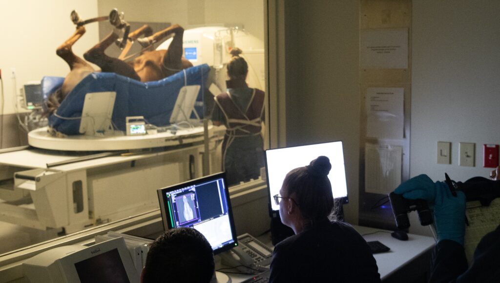
Fetal alcohol syndrome-related craniofacial differences could be seen in offspring born to fathers who regularly consumed alcohol to the legal limit.
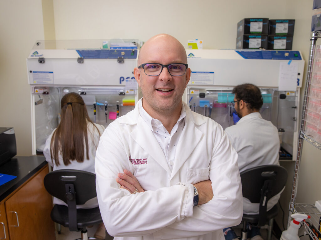
Dr. Michael Golding
According to the U.S. Surgeon General, women should not drink alcoholic beverages during pregnancy because of the risk of birth defects in their unborn child. However, research at Texas A&M University now demonstrates that a father’s alcohol consumption also links to growth defects that affect the development of his offspring’s brain, skull, and face.
However, because men drink more and are more likely to binge drink than women, Dr. Michael Golding, an associate professor in the School of Veterinary Medicine & Biomedical Sciences’ Department of Veterinary Physiology & Pharmacology, and his team set out to challenge the existing dogma, using a mouse model to examine what happens when the mother, father, and both parents consume alcohol.
In a new article published in the Journal of Clinical Investigation, Golding and his team found that male alcohol consumption before conception caused FAS brain and facial growth defects.
“We found that male exposures actually drive certain craniofacial differences much stronger than maternal exposures do, so this programming effect that’s coming through sperm has a profound effect on the organization of the face and the growth and proportion of different facial features,” Golding said. “When it was the dad drinking, we saw a profound shift in the organization of the face.”
FAS is hard to diagnose, but when doing so, doctors currently look for abnormal facial features; lower-than-average weight, height, or both; central nervous system problems such as a small head size, problems with attention and hyperactivity, or poor coordination; and verification of maternal alcohol use during pregnancy.
“When doctors suspect a child has FAS, they sit down with the mother to confirm the diagnosis by discussing her drinking habits during pregnancy,” Golding shared. “It’s not uncommon for the mother to deny consuming alcohol while pregnant. When they do, there’s this stigma or this notion that women are lying about their alcohol use.”
Golding said this research, which was funded by a Medical Research Grant from the W.M. Keck Foundation and the NIH National Institute on Alcohol Abuse and Alcoholism, reveals a potential blind spot in the current diagnostic criteria for FAS, the most severe form of Fetal Alcohol Spectrum Disorder (FASD), which requires documentation of maternal alcohol use during pregnancy.
“Our research proves there’s a plausible alternative explanation — the father’s contribution, which has never been examined before,” he said. “In this study, we call into question the dismissal of the mother’s denial and really examine the capacity of male alcohol use to induce FAS growth defects.”
Golding explained that findings from his holistic approach that examines both parents’ contributions to FAS reveal the need for two critical changes.
“First is the recognition of the importance of male health in pregnancy outcomes and fetal health,” he said.
Golding pointed out that paternal health before conception is a novel consideration in terms of pregnancy outcomes and fetal health; as a result, raising awareness of the role a father’s health plays in his offspring’s health is just as important as awareness of the mother’s contributions from preconception and through gestation.
“Research examining fetal health is overwhelmingly focused on maternal health,” he said. “I’m not saying that this is not appropriate; I’m just saying it’s not the complete picture and we need some balance.”
“The second,” he said, “is the fact that both parents are responsible for preventing alcohol-related birth defects.”
FAS has significant, life-changing consequences for children. Because their study identified FAS-related craniofacial differences in offspring born to fathers who regularly consumed alcohol to the legal limit, Golding pointed out that both parents should commit to limiting or omitting their alcohol consumption before trying to become pregnant.
Ultimately, Golding emphasizes that the first step in this process is expanding messaging outreach about the reproductive dangers of alcohol use to both parents.
“Change the alcohol warning label to remove the maternal emphasis and put it on both parents to say, ‘The decision to consume this beverage can have significant, life-changing consequences to a future child,’” he said. “Right now, the warning label only conveys part of the story. We must get that message out into the world as quickly as possible.”
###
For more information about the Texas A&M College of Veterinary Medicine & Biomedical Sciences, please visit our website at vetmed.tamu.edu or join us on Facebook, Instagram, and Twitter.
Contact Information: Jennifer Gauntt, Director of VMBS Communications, Texas A&M College of Veterinary Medicine & Biomedical Sciences, jgauntt@cvm.tamu.edu,979-862-4216
While on a walk at Lake Bryan, a couple’s dog encountered a box and became excited after smelling something inside. Curious to see what was causing their dog to react, to their surprise, they found a puppy inside the box.
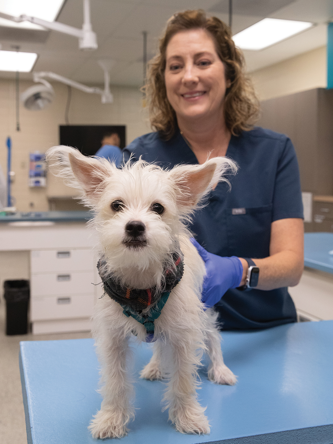
Immediately, the couple brought the puppy, who would soon be known as Gertie, to Texas A&M’s Small Animal Teaching Hospital (SATH). It was unclear how long the small, white terrier-mix was left alone in the box, but she appeared to have broken bones and was covered in fleas, dehydrated, and malnourished.
After Gertie’s initial examination, the couple, despite having no intentions of keeping her, brought Gertie back to the SATH for an orthopedic appointment with Dr. Laura Peycke, a clinical associate professor at the Texas A&M College of Veterinary Medicine & Biomedical Sciences (VMBS).
At the appointment, Peycke and a veterinary technician, Wendy Greathouse, schemed to bring together Gertie and Dana Whitaker, an SATH assistant hospital administrator. Whitaker had had a running joke that she would eventually get a “scruffy purse dog,” and Peycke, in introducing her to Gertie during the examination, made that joke a reality.
Meeting For The First Time
While in the SATH emergency room during her initial visit, radiographs revealed that two of the most concerning injuries Gertie sustained included a partially healed left femur fracture and partially healed right pelvic fracture.
“I think someone stepped on her and they panicked because they knew she was broken and discarded her,” Whitaker said.
Specialty appointments at the SATH can be hard to schedule quickly because they are typically booked months in advance, but Gertie was able to get an orthopedic appointment with Peycke only two days after her first ER visit to have her fractures examined and to ensure she would not need surgery to repair the fractures.
“When Dr. Peycke saw this little thing in her orthopedic appointment and found out the people who brought her in were not planning on keeping her, she and Wendy plotted together to get me in Gertie’s path,” Whitaker said.
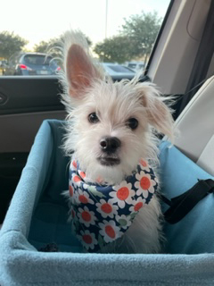
Walking to the exam room, Whitaker wasn’t sure if she’d keep Gertie but upon meeting her, she couldn’t resist taking Gertie home to ensure she was cared for throughout her healing process.
“When I met Gertie, everything clicked,” Whitaker shared. “Gertie needed to end up with me, and she’s mine now.”
Since then, Whitaker and Gertie have been a team on the road to recovery.
A Healthy Puppy Means A Happy Puppy
Gertie’s journey to becoming a happy, healthy puppy was off to a good start now that she had Whitaker and the SATH veterinary team looking after her.
Gertie required surgery in January to repair an inguinal hernia, a tear in the body wall that would allow organs to pop out of their place underneath the skin, and her fractures have since improved.
“Gertie’s fractures are now healed, but not in the place we would want them to be healed if we were in control of her recovery immediately after they occurred,” Whitaker said. “If we saw her the day she was injured, we could have fixed things.”
Because these fractures were not properly taken care of when they occurred, Gertie developed quadriceps contracture, the physical shortening of muscle length that leads to stiff muscles and prevents stretching, full range of motion, and joint mobility in her left leg.
Even though her left leg does not function like her other legs and is only used for balance when necessary, her daily activities of running and playing are not disrupted, allowing Gertie to expel her puppy energy.
“When she runs, she runs fast and her left hind leg, the one that was fractured, tags along with her,” Whitaker explained. “Because she has quadriceps contracture, there is no way to recover the muscle because it’s bound up so tight. There’s not anything we can do, but it doesn’t cause her any pain.”
There is concern, though, that as Gertie ages, the head of the femur and the pelvis it sits in will rub together when the cartilage coating the two bones wears down, causing Gertie pain.
“If she needs any orthopedic work in the future, we’ll probably have to do a femoral head ostectomy,” Whitaker said. “We would cut the head of the femur off, so the muscle would be holding everything together, and there’s no bone sitting in the pelvis.”
But for now, Gertie is a pain-free, energetic, and happy 8- or 9-month-old puppy, whose favorite pastime is chasing balls and toys and bonding with other furry visitors during her adventures visiting the SATH with Whitaker.
“She is a hundred percent mischievous and just a happy-go-lucky pup,” Whitaker shared. “I look forward to seeing how her personality will grow and settle as she becomes an adult.”
###
For more information about the Texas A&M College of Veterinary Medicine & Biomedical Sciences, please visit our website at vetmed.tamu.edu or join us on Facebook, Instagram, and Twitter.
Contact Information: Jennifer Gauntt, Director of VMBS Communications, Texas A&M College of Veterinary Medicine & Biomedical Sciences, jgauntt@cvm.tamu.edu,979-862-4216
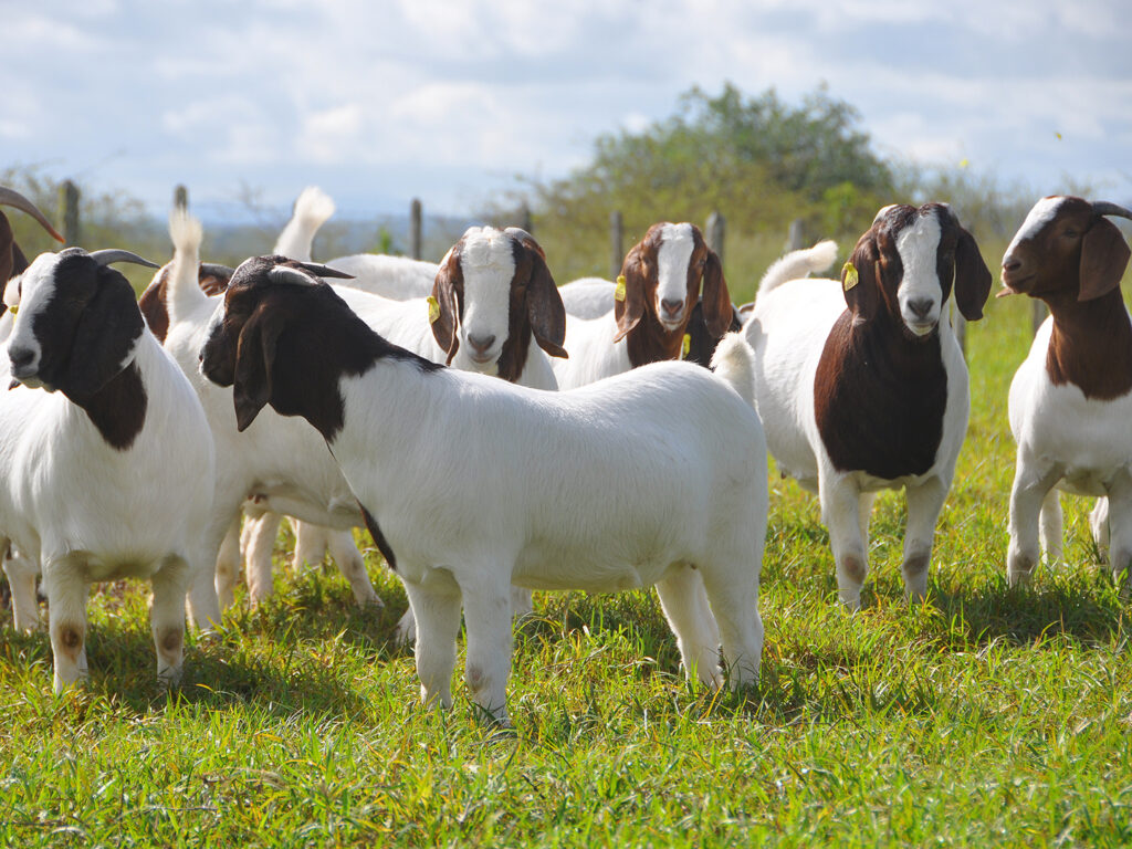
Urinary stones, or solid masses made from minerals in the urine, develop for a variety of reasons and can be an extremely painful experience for humans and pets alike. Maintaining a healthy lifestyle can prevent stones from developing, which is especially beneficial for male goats, who commonly experience this condition.
Dr. Shannon Reed, a clinical associate professor in food animal medicine at the Texas A&M School of Veterinary Medicine & Biomedical Sciences, says urolithiasis — or the formation of stones in the urinary tract — should always be treated by veterinarians since home remedies often delay and complicate the recovery process.
“Suspicion of urolithiasis, or a ‘blocked goat,’ should always warrant seeing a veterinarian, as animals that cannot urinate experience life-threatening complications rapidly,” Reed said. “Delaying evaluation and treatment will both threaten the animal’s health and increase the cost associated with treatment.”
A goat impacted by urolithiasis will show signs like attempting to urinate repeatedly without actually urinating; bleating with a flagging tail, which is a tail that is held stiff and high while moving back and forth; appetite loss; and an outwardly swollen belly.
Once owners take their goats to a practitioner, Reed explained that a veterinarian will do a complete physical examination and often sedate the animal to examine for stones; additional information, such as age and where in the urinary tract the stone is stuck, can determine the next steps.
“Young animals under 8 months are more likely to have stones that are easier to resolve and will dissolve with treatment, while animals with hard stones, called calcium carbonate stones, will need surgery to resolve the issue,” Reed said. “Animals might also need intensive care if their kidneys are affected by the urine blockage or need an alternate way to pass urine, such as a tube inserted into their bladder.”
Stones can be exacerbated by inappropriate diet, poor water consumption, and poor water quality, according to Reed.
But because urolithiasis is complicated by obesity, Reed advises owners to first provide goats with a low-grain diet that includes a mineral block or loose mineral supplements as a way to prevent stones from forming.
“Animals that are fed too much grain are more likely to get urinary blockages, so most goats do not need grain if they are fed high-quality grass hay with a mineral source and are not growing, pregnant, or lactating,” Reed said. “Since most adult goats also need a mineral supplement beyond hay to balance their diet but do not need extra calories, it is better to offer a mineral block.”
Since diets can vary from goat to goat, Reed suggests owners check with their veterinarian that their goats are meeting their dietary requirements.
Reed also encourages owners to continually maintain fresh, clean water for goats to increase a goat’s water consumption.
“Increasing water consumption increases urine production, so goats will pee more often, leading to less sediment gathering in the bladder to form stones,” Reed explained. “In warmer months, high water temperature and algae will decrease water consumption, so owners should place water in the shade and clean water buckets frequently; using heated water buckets in the wintertime can also be helpful.”
Hard water, or water that is high in minerals, can contribute to stone formation as well. Because of this, owners should filter water from a hose or from other sources such as wells so that the water’s quality is improved. Reed recommends owners buy a filter that can attach to the end of the hose as an inexpensive yet effective way to filter water.
With an appropriate diet, additional minerals, and fresh, clean water, goats can be protected against urinary stones. If they do experience urolithiasis, goats should always be taken to their veterinarian for treatment so that they can return to their daily life sooner.
Pet Talk is a service of the College of Veterinary Medicine & Biomedical Sciences, Texas A&M University. Stories can be viewed on the web at vetmed.tamu.edu/news/pet-talk. Suggestions for future topics may be directed to vmbs-editor@tamu.edu.
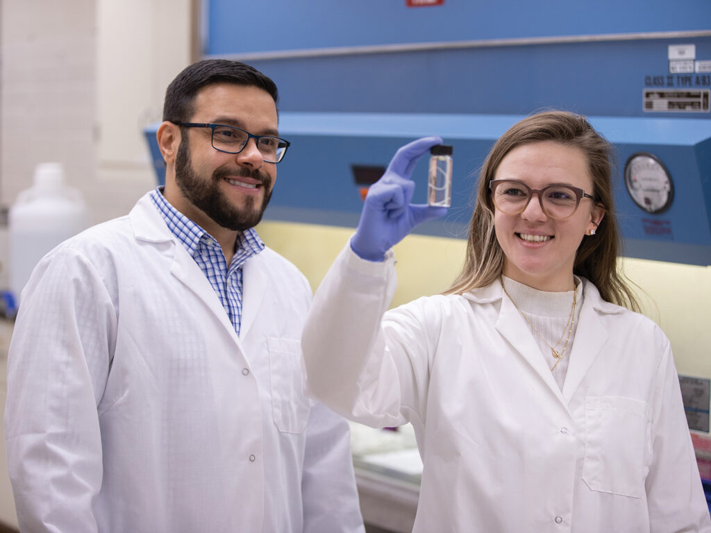
Dr. Guilherme Verocai and former postdoctoral researcher Dr. Caroline Sobotyk de Oliveira
While many veterinarians will admit to having a favorite animal, worms are usually not at the top of the list.
For Dr. Guilherme Verocai, however, worms—specifically those that live parasitically inside another organism—are the most fascinating animals on the planet.
“Worms are my life,” said Verocai, a clinical assistant professor in the Texas A&M School of Veterinary Medicine & Biomedical Sciences’ (VMBS) Department of Veterinary Pathobiology (VTPB). “They’re pretty important, of course, in the medical and veterinary fields. But beyond that, they’re diverse and complex.
“They’re animals living in, on, or off another animal,” he continued. “They’re much more intelligent than viruses and bacteria. Evolutionarily speaking, they’re very evolved organisms; they had to adapt to this parasitic lifestyle, which, arguably, is the most successful lifestyle on Earth.”
Verocai’s fascination with parasitic worms, also known as helminths, has influenced his entire career, which has taken him from Brazil to Canada to Africa and, finally, to Texas A&M, where he keeps a busy schedule as an educator, researcher, and diagnostician.
Finding A Passion
Helminths first caught Verocai’s attention during his first year of veterinary school at the Universidade Federal Rural do Rio de Janeiro in Brazil.
“Early in vet school, I had courses in parasitology and I fell in love, basically,” he said. “I started doing parasitology research and I was a teaching assistant in parasitology, and I never left the research and teaching aspects.”
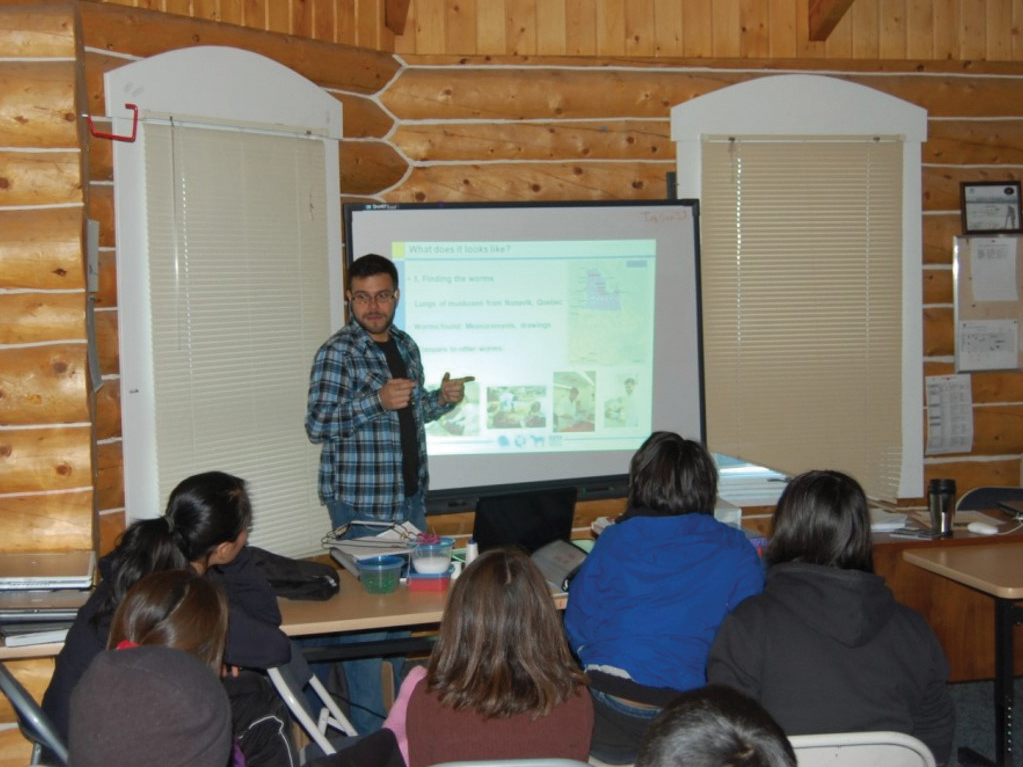
Verocai teaching in Canada
After earning his Doctor of Veterinary Medicine (DVM) degree in 2005 and a Master of Science degree in veterinary parasitology in 2008, Verocai decided to expand his horizons, quite literally, by enrolling in a Ph.D. program in Canada.
At the University of Calgary, he chose to pursue a secondary interest in wildlife medicine—while still keeping his focus on worms, of course.
“I ended up in a very nice university at the right time,” he said. “The graduate program in veterinary medicine at the University of Calgary was brand new, so there were a lot of excited professors coming in full of ideas, as well as a lot of grad students coming from a bunch of different places on Earth. It was a pretty fun, diverse, and inspiring place to be.”
Verocai’s research project there focused on discovering a new species of lungworm that scientists believed to be living in large mammals above the Arctic Circle.
“Before I got there, they had found larvae in feces of caribou, moose, and musk oxen, and we knew it was something new because they didn’t match the genetics of any related worms,” Verocai said. “I really wanted to discover one of the adults and describe this new species, and I had no doubt that I would find them.”
Native hunters, including the Inuit and Dene ethnic groups, sent the Canadian research team samples of lung tissue from caribou and musk oxen to search for adult specimens of the new species.
“It was like working in the dark because the worms didn’t cause any obvious pathology or any lesions in the lungs,” Verocai said. “We were cutting through, washing, and sieving the tissue samples to look for the hairlike worms, which were only a few centimeters long. It was a big moment when we found the first adult worms in the lungs of musk oxen from Northern Quebec.”
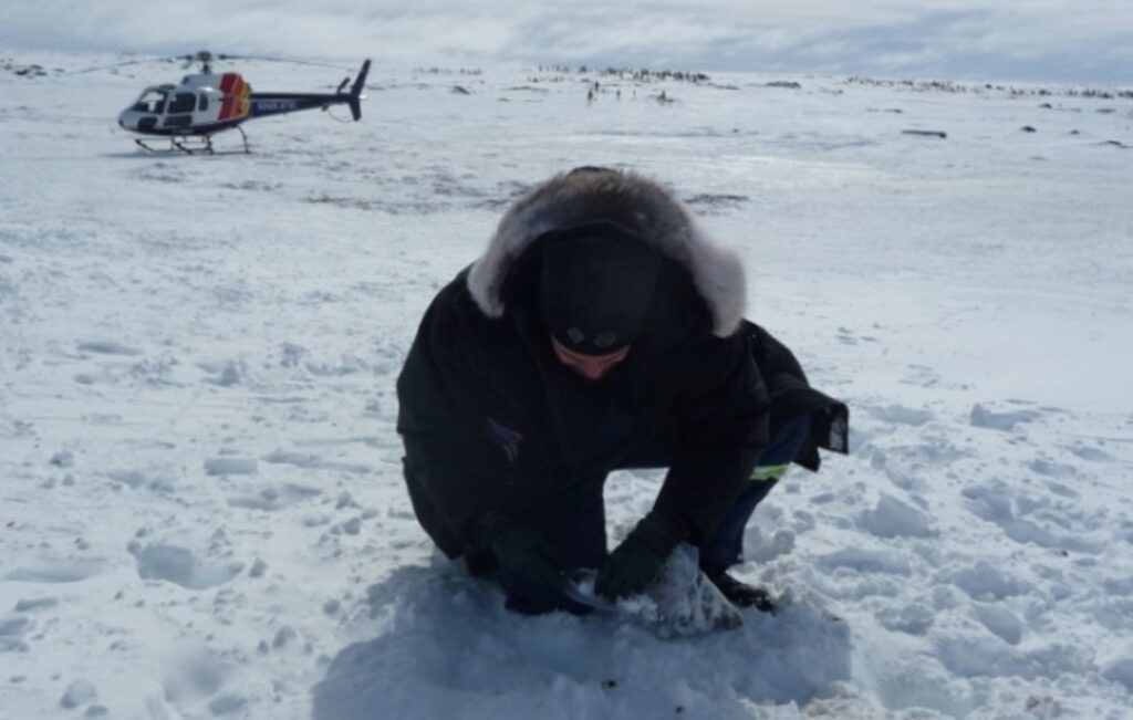
Verocai collecting samples in the Arctic
During the project, Verocai also spent a lot of time in Arctic regions, talking to native peoples and collecting samples.
“I’m from Rio de Janeiro, a beach town, and here I was collecting poop by helicopter and chasing musk oxen and caribou in sub-Arctic and Arctic areas,” he said with a laugh.
“We went from village to village, talking about veterinary medicine, wildlife health, and the importance of parasites,” he said. “Everything was being translated into their native language. It was a pretty interesting experience.”
When it came time to name the new species, Verocai’s team decided to credit the Sahtu Region of Canada’s Northwest Territories, where the lungworm larvae were first detected.
“We wanted to name it something meaningful for them in their native language because of the importance of caribou in their culture,” Verocai said. “In North Slavey, one of the dialects for the Dene people, they call the Arctic ‘elegu nene,’ or ‘cold land.’ Varestrongylus is the genus of the parasite, so we named it ‘Varestrongylus from the cold land,’ or Varestrongylus eleguneniensis.”
Back To Warmer Weather
Once Verocai earned his Ph.D. in 2015, he moved south to begin working as a postdoctoral researcher in the University of South Florida’s Department of Global Health, where he could study a pair of related parasites that he found especially interesting.
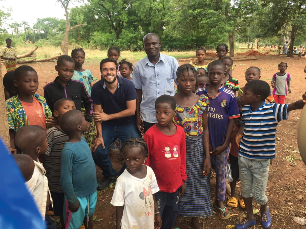
His main project, funded by the Bill & Melinda Gates Foundation, involved traveling to the West African country of Burkina Faso to study a nematode called Onchocerca volvulus that causes onchocerciasis, also known as river blindness, in people. This disease is one of the World Health Organization’s (WHO) top 20 neglected tropical diseases.
Verocai in Burkina Faso
Verocai also went back to his veterinary roots and spent his spare time studying Onchocerca lupi, an obscure zoonotic parasite that causes a similar disease in dogs and cats.
He has continued to study O. lupi since he began his career at Texas A&M in November 2018; to this day, those searching the parasite on Google will find Verocai’s publications as two of the top three results.
“Parasites are a neglected part of biodiversity because, well, it’s tough to study them,” Verocai said. “Internal parasites, especially, are even more challenging to work with than external ones like fleas and ticks.”
Continuing to rise to this challenge, Verocai has taken on several new projects at the VMBS, including studying Dracunculus medinensis, also known as the Guinea worm.
The Guinea worm is similar to O. volvulus in that this nematode also causes one of the WHO’S top 20 neglected tropical diseases, dracunculiasis (Guinea worm disease). The project is also funded by another well-known organization, The Carter Center.
“The scientific community once thought it was strictly a human parasite, but then we realized that dogs; to a lesser extent, cats; and, in some countries like Ethiopia, baboons were acting as reservoirs and helping cycle the parasite,” Verocai said.
While the Guinea worm does not usually kill its host, it can cause blindness, blisters, and swelling, all of which negatively impact an individual’s livelihood, both physically and socially.
“Parasites are trying to survive like all of us and, most of the time, killing the host is not an ideal strategy for them to continue on,” Verocai said. “But there is still a big impact on health and general well-being, as well as an economic burden on the infected humans.”
This aspect of parasites translates to animal hosts as well, including pets and food animals.
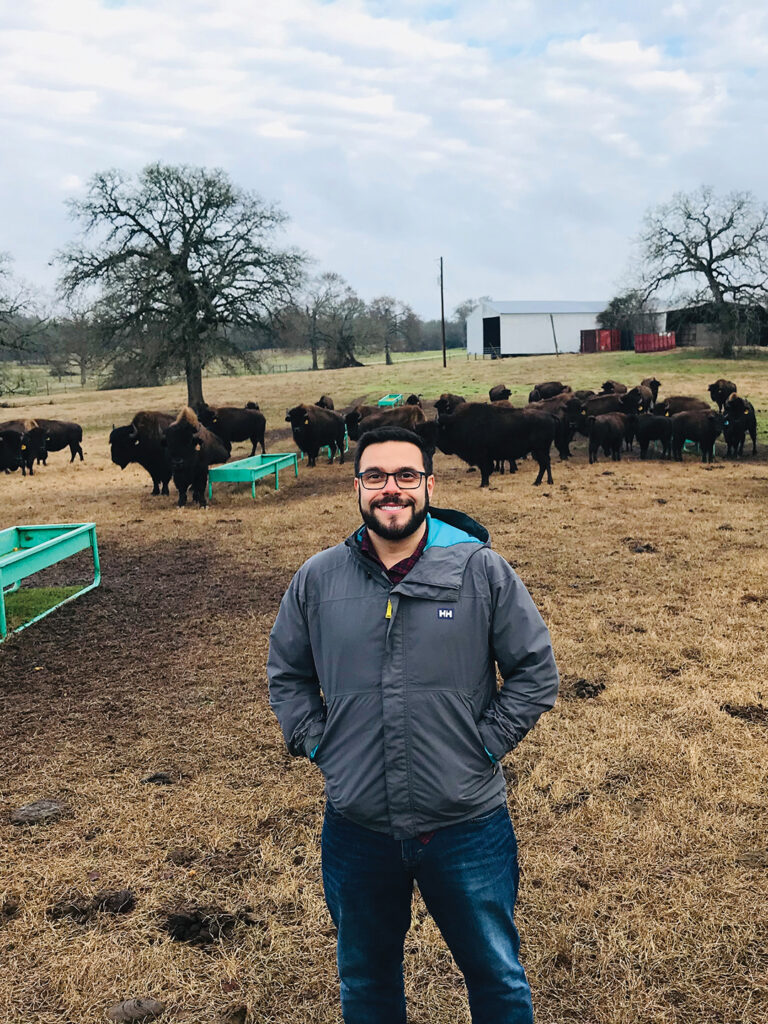
Verocai at a bison ranch in Texas
“That’s what a lot of parasites will do with our pets—the hosts are not dying, but they could be healthier; they’re not living their best life,” Verocai said. “Thinking about animal production, every infected animal is producing less than it could. That’s billions of dollars that go to waste because of parasites impacting production animals.”
Because of the far-reaching negative impacts of parasites on animals, one of Verocai’s roles at the VMBS is directing the Parasitology Diagnostic Laboratory, which helps veterinarians diagnose parasitic infections in companion animals, exotics, livestock, and wildlife.
“Diagnostics is a big portion of my appointment here,” he said. “It’s figuring out which parasites are present, plus triaging the biodiversity of parasites and figuring out what we don’t know. We will only know how to properly deal with things that we know about.”
These diagnostic services tie in with his research mission; one of Verocai’s current projects involves looking for new diagnostic markers for heartworms, a mosquito-borne parasite of dogs, and other helminths of veterinary and medical importance.
“Diagnostic markers, which are secretory/excretory products of the parasites, will be found in the organism they’re inside, circulating in biofluids like blood,” he said. “Right now, there are diagnostic markers for cancer and various other human diseases, but they are less explored in parasitology and veterinary medicine. There’s a whole world out there to be studied.
“We’ve had projects with heartworms and other filarial worms, trying to figure out which microRNAs they are excreting/secreting,” he said. “Then, for the Guinea worm, we’re adapting and expanding what we were doing for heartworms in a more global health direction. Ideally, we can use the same test to screen samples from any host species.”
Training The Next Generation
One final aspect of Verocai’s role at the VMBS is educating and mentoring students, from the veterinary students enrolled in his Agents of Disease courses to the graduate students and postdocs working in his lab.
“Teaching and mentoring are very rewarding,” Verocai said. “I’m trying to convince everybody that parasites are the most amazing things on Earth. Even though I might not be convincing enough, showing how much I care shows that parasites matter.”
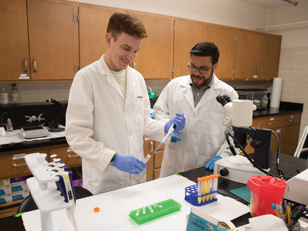
Verocai and recent DVM graduate Andrew Brown
During the summer of 2021, as a mentor in the VMBS’ Veterinary Medical Scientist Research Training Program (VMSRTP), Verocai spent 14 weeks working with third-year veterinary student Hannah Danks to characterize a new species of lungworm in North American bison.
Recent research by the VMBS’ Dr. James Derr suggests that all bison in North America have some cattle DNA, so a logical assumption would be that the two species share the same parasites as well.
“Bison almost went extinct. How could they keep cycling their own parasites?” Verocai asked. “But when we looked at samples from different herds, they were not the same as the cattle lungworm. It seems that the bison lungworms are a success story of their own, a parasite that, like bison, survived from the brink of extinction.”
Verocai’s continued promotion of parasitology among Texas A&M’s DVM and graduate students and parasitology residents has already resulted in the formation of the Texas A&M Student Chapter of the American Association of Veterinary Parasitology, which he co-advises with clinical assistant professor Dr. Meriam Saleh.
“We’re trying to fill the gaps and make sure parasitology is there as a career opportunity,” he said. “Did I know I would be a parasitologist when I got into vet school? Of course not. If students do not perceive it as an option, it’s never an option.
“This is why I also have a commitment to diversity,” he continued. “I happen to be gay, I happen to be Brazilian, and that’s part of who I am. It matters. You need to see diversity to be able to respect it. It’s all about finding that passion.”
For more information about the Texas A&M College of Veterinary Medicine & Biomedical Sciences, please visit our website at vetmed.tamu.edu or join us on Facebook, Instagram, and Twitter.
Contact Information: Jennifer Gauntt, Director of VMBS Communications, Texas A&M College of Veterinary Medicine & Biomedical Sciences; jgauntt@cvm.tamu.edu; 979-862-4216
Humans typically avoid raw foods because of the risk of developing a salmonella infection, a common bacterial disease that harms the intestinal tract. While humans can be infected with salmonella more easily than animals, there is still a risk of pets developing the infection as well.
Dr. Katie Tolbert, a clinical associate professor in small animal and comparative gastroenterology at the Texas A&M School of Veterinary Medicine & Biomedical Sciences, says that while some species of salmonella cause symptoms that are self-limiting — meaning, those symptoms will resolve on their own — salmonella is not something that should be taken lightly, as certain species can cause illness and disease if ingested.
“Salmonella is a bacteria frequently found in the gastrointestinal tract of many mammals and birds, but some species of salmonella produce toxins that cause infections when the bacteria adheres to the lining of the gut, causing gut inflammation and damage,” Tolbert explained. “The infection can travel to other sites of the body, such as the joints or heart, and if the infection is not recognized, it could lead to severe consequences, including death.”
Tolbert pointed out that pets who are infected with toxin-producing species of salmonella are generally those who have ingested raw foods.
“Most raw foods that a pet would eat would be fed to them by their owner or via scavenging through household trash, although it is possible for unsupervised pets to have access to animal carcasses that could harbor bacterial infections,” Tolbert said.
If a pet displays significant symptoms of a gastrointestinal illness, Tolbert strongly advises owners to visit their veterinarian as soon as possible to determine an appropriate treatment plan and if the illness is the result of a salmonella infection.
“Warning signs that should prompt a pet owner to bring their dog or cat to be evaluated include prolonged reluctance to eat for longer than 24 to 48 hours; fever; persistent vomiting or diarrhea; lethargy for longer than 24 hours; signs of dehydration, such as dry or tacky gums; and signs of pain, shown through abnormal behaviors such as hiding or growling,” Tolbert explained.
Veterinarians may hospitalize pets and provide them with intravenous fluids and supportive care if the illness is severe.
The most common symptom of a mild form of salmonella infection in pets, however, is diarrhea that will often resolve on its own, whether treatment is involved or not.
But pets can have diarrhea for different reasons, so Tolbert explained that it can be difficult to know if the diarrhea is caused by a salmonella infection, bacterial infection, or noninfectious causes. Noninfectious causes include pancreatitis — or the inflammation of the pancreas — and dietary indiscretions — the tendency for animals to eat items outside of their normal diets, such as household trash.
“Since self-limiting diarrhea resolves with supportive care, identifying the specific cause of diarrhea may not be pursued unless there is a known risk for salmonella infections, which would be harmful for the pet and other members of the household,” Tolbert said.
If the veterinarian chooses not to identify the cause of the diarrhea, Tolbert encourages owners to provide supportive care by keeping pets well hydrated and feeding them highly digestible diets, which are foods and nutrients that pets can easily digest and absorb, as well as by following your veterinarian’s dietary recommendations.
“Most commercial diets are highly digestible, but there are also specialized, therapeutic diets available through a veterinarian’s office and are used for both acute and chronic gastrointestinal diseases,” Tolbert said. “These specialized diets are highly digestible but also contain ingredients that are designed to help alleviate vomiting and diarrhea, such as fibers, absorbents, and antioxidants.”
Nevertheless, owners should visit their veterinarian if they suspect a pet’s diarrhea is caused by their pet ingesting raw foods because pets experiencing mild salmonella infection symptoms can shed the bacteria and infect other pets or humans, who may develop a more severe infection. Owners should also ask their veterinarian if isolation is necessary to prevent the bacteria from spreading.
Additionally, Tolbert recommends owners always practice good hand and environmental hygiene, as this can prevent the infection from easily spreading between humans and pets.
Owners should be aware of the causes, symptoms, and treatments in the event their cat or dog develops a salmonella infection. Treating salmonella infections sooner rather than later can help your furry friend recover quicker and keep the rest of the family in good health too.
Pet Talk is a service of the School of Veterinary Medicine & Biomedical Sciences, Texas A&M University. Stories can be viewed on the web at vetmed.tamu.edu/news/pet-talk. Suggestions for future topics may be directed to vmbs-editor@tamu.edu.
Texas A&M is also the most highly ranked among all Southeastern Conference (SEC) universities with veterinary schools.
The VMBS’ 2023 rankings took a large leap up from 2022, when the VMBS was ranked No. 20 worldwide. The annually compiled rankings cover a total of 54 disciplines, grouped into five broad subject areas.
“We are delighted by this recognition, and we are honored to represent Texas A&M University, the Texas A&M System, and the state of Texas in the QS World University Rankings,” said Dr. John August, the Carl B. King Dean of Veterinary Medicine at Texas A&M. “This achievement was made possible by our outstanding faculty, students, and staff who do remarkable work each day at the VMBS to fulfill our teaching, research, and patient care missions. It is always rewarding to see their collective hard work recognized, especially on a global and national scale.”
For more than 100 years, the VMBS has been dedicated to enhancing animal and human health through transformational education, discovery and innovation, patient care, and public service that impact our diverse and evolving world.
Transformational education is at the core of the VMBS’ existence; this is evident in the fact that the VMBS is one of the largest Doctor of Veterinary Medicine (DVM) training programs in the country, with a class size of 180. In addition, the Texas A&M DVM Class of 2023’s North American Veterinary Licensing Examination first-attempt pass rate was 93%, compared to the national average of 79%.
The VMTH has the distinction of being Texas A&M’s largest educational laboratory and the state’s only veterinary medical teaching hospital. DVM students completing their fourth-year clinical rotations through the VMBS see almost 30,000 animal cases annually from across North America and receive training in more than two dozen large and small animal veterinary specialties.
VMBS faculty and students also turn basic science into novel discoveries that set the course for improving human and animal health; publication citations and the h index, which reflects the productivity and impact of a university’s basic science and clinical research activities, were among the criteria assessed in the QS rankings.
“This ranking reflects the high-quality and impact of the translational and basic biomedical research being conducted at our school by our world-class faculty and the students they mentor,” said Dr. Ramesh Vemulapalli, VMBS executive associate dean.
The VMBS’ Research & Graduate Studies Office administers one of the largest graduate programs at a school of veterinary medicine, and veterinary students gain exposure to research through programs such as the Veterinary Medical Scientist Research Training Program and through clinical trials offered within the VMTH.
In addition, through the VMBS’ Veterinary Education, Research, & Outreach (VERO) initiative, housed on the West Texas A&M University campus in Canyon, veterinary students get exposure to large animal and mixed animal medicine through VERO partnerships with Texas’ thriving livestock and food animal industries as well as with rural veterinary practitioners.
The VMBS’ 2+2 DVM program, launched in 2021, offers up to 18 students the opportunity to complete the first two years of their veterinary medical education at VERO before joining their peers in College Station for their third and clinical years. VERO also offers opportunities for DVM students to return to the Texas Panhandle during fourth-year clinical rotations in dairy practice, feedlot medicine/surgery, cow-calf practice, and rural/mixed animal general practice.
QS World University Rankings are used by prospective students to help identify the leading universities in a particular subject. Universities are ranked based on journal article citations and by the results of major global surveys of employers and academics.
###
For more information about the Texas A&M College of Veterinary Medicine & Biomedical Sciences, please visit our website at vetmed.tamu.edu or join us on Facebook, Instagram, and Twitter.
Contact Information: Jennifer Gauntt, Director of VMBS Communications, Texas A&M College of Veterinary Medicine & Biomedical Sciences, jgauntt@cvm.tamu.edu,979-862-4216
Three faculty members at the Texas A&M School of Veterinary Medicine & Biomedical Sciences (VMBS) were among the 25 recipients from across Texas A&M University to be recognized with 2023 Distinguished Achievement Awards by The Association of Former Students.
The university-level awards honor faculty and staff who exhibit the highest standards of excellence and are held in high esteem by their colleagues.
These awards are among the most prestigious available to faculty and staff members at Texas A&M. Recipients are chosen by a campuswide committee of faculty, staff, students, and former students.
The recipients will be honored with a commemorative plaque and engraved watch during an awards luncheon on April 24 in the Memorial Student Center’s Bethancourt Ballroom.
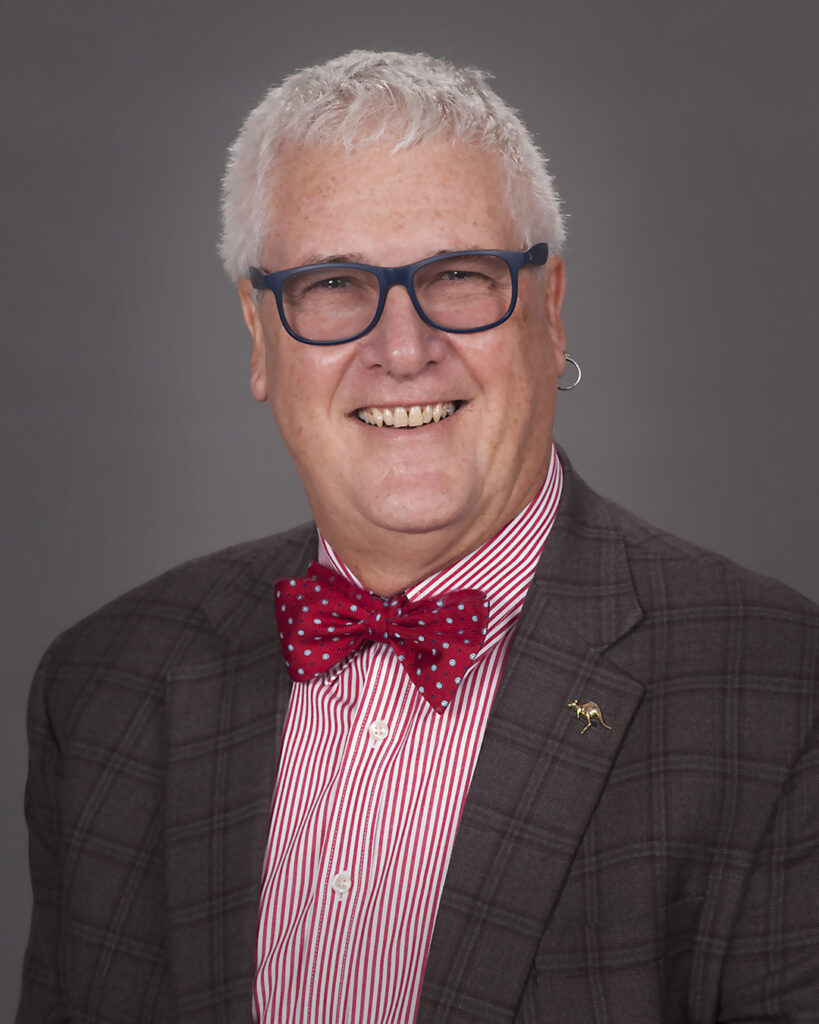
Dr. Larry J. Suva
Dr. Larry Suva
Suva is a leading expert on the skeletal consequences of disease, with research interests including breast cancer bone metastasis, multiple myeloma, fracture healing, bone regeneration, bone anabolism, and the investigation of rare bone diseases and bone infections.
“A feature of his work throughout his career is a willingness and eagerness to take on difficult and important topics of clinical and social relevance,” said one nominator. “Dr. Suva’s career and research continue in a steep trajectory, the most recent example of which is the work he has had funded by the NIH to study fracture healing in the context of the bone disease in Down’s Syndrome.”
Some of Suva’s most important research contributions have been the discovery of human parathyroid hormone‐related protein (PTHrP) and the hormonal regulation of bone metastasis.
“Dr. Suva is considered one of the world’s foremost experts on PTHrP,” the nominator said. “He is one of the most recognized and respected scientists in this very important field. The impact on the treatment of osteoporosis because of Dr. Suva’s seminal discovery is truly remarkable, and Texas A&M University is fortunate to have recruited such an exceptional scientist to join its ranks.”
Suva joined the VMBS in 2015 and is dedicated to advancing research, graduate student success, and diversity, equity, and inclusion at the school.
“Dr. Suva’s life is characterized by enthusiasm for research and devotion to science. He nurtures the same enthusiasm in his trainees and he creates an environment in which they can succeed,” the nominator said. “All of the graduate students and postdoctoral fellows who have had the privilege of receiving education and training from Dr. Suva have gone on to successful careers in academia, private practice, government agencies, or large corporations.”
Prior to joining the VMBS, Suva received his Ph.D. in medicine from the University of Melbourne (Australia), was a postdoctoral researcher at Merck, and was a faculty member at Harvard Medical School.
Dr. Johanna Heseltine
Dr. Johanna Heseltine
Heseltine plays an important role in veterinary students’ preclinical education and in their clinical year at the Texas A&M Veterinary Medical Teaching Hospital. Her courses generally focus on the urinary tract, respiratory system, and other aspects of internal medicine.
“Dr. Heseltine is really an amazing mentor and I look up to her a great deal,” said one of Heseltine’s former students. “Her willingness and enthusiasm to teach made me eager to learn and fueled my passion for veterinary medicine. I appreciate that she was available to us for questions about cases but also for general life questions.”
Heseltine’s courses rely on student participation and increasingly complex real-world scenarios. She also was a vital member of the faculty team that revised Texas A&M’s veterinary curriculum in 2017.
“She has contributed to curricular development, created novel educational modules, generates scholarship focused on teaching and learning, and is strongly focused on facilitating the development of lifelong learners,” one nominator said. “She has been recognized several times for her excellence as a teacher, through the Richard H. Davis Award (2016 and 2019), Juan Carlos Robles Emanuelli Teaching Award (2018), the John Miliff Award for Teaching (2020), and an AFS school-level Teaching Award (2022).”
In addition to training veterinary students, Heseltine is an educator and mentor for interns and residents in the Small Animal Teaching Hospital’s Internal Medicine Service.
“Dr. Heseltine must act as a role model, helping students and residents navigate emotionally charged clients and challenging cases,” the nominator said. “She helps her trainees acquire complex physical skills such as heart and lung auscultation as well as interpretation of diagnostics such as ultrasounds and blood work. Simultaneously, Dr. Heseltine helps her students and residents understand best practices in medical decision making and provides clear, professional feedback.”
Heseltine joined the VMBS’ Department of Small Animal Clinical Sciences in 2014 after earning her DVM degree from the University of Saskatchewan in 1998, attaining diplomate status from the American College of Veterinary Internal Medicine in 2004, and spending several years in private practice and other academic teaching positions.
Dr. Anton Hoffman
Dr. Anton Hoffman
Hoffman has been a member of the VMBS’ Department of Veterinary Integrative Biosciences for 35 years. He has contributed substantially to the education of both veterinary and graduate students.
“Dr. Anton Hoffman embodies what it means to be a superior teacher,” one nominator said. “He is not satisfied with the status quo or teaching a set of facts. Instead, Dr. Hoffman thinks outside that box and works to continually improve his teaching and approach to class in order to provide students the most meaningful and long-lasting learning experiences as possible.”
His courses focus on small and large animal anatomy, physiology, organ dysfunction, and professional and clinical skills. In addition to classroom and laboratory teaching, Hoffman has served as a mentor for many DVM students, including two who have gone on to become VMBS faculty members themselves.
“From the moment I met him, it was obvious to me that education was a priority and teaching anatomy was a passion of his,” said one of Hoffman’s former students. “One of Dr. Hoffman’s most attributing qualities is his research to deliver creative, thought-provoking, and appropriate lessons that match all students’ abilities. For example, Dr. Hoffman used illustrations and life-sized models to help us understand the intricacies that lie within anatomy.”
Some of Hoffman’s past awards and recognitions include the Presidential Professor for Teaching Excellence Award in 2011, the Zoetis Distinguished Veterinary Teaching Award in 2014, the National Pfizer Distinguished Veterinary Teaching Award in 2011, the Chancellor’s SLATE Award in 2009, and twice being selected as a qualifying examination question writer for the National Board of Veterinary Medical Examiners.
“One of the attributes that makes Dr. Hoffman truly amazing as an instructor is his ability to connect with his students and engage them,” the nominator said. “He fosters an atmosphere that ensures students know that they can always go to him whether it is a problem they are having with course content or in their personal lives.”
Hoffman received his DVM degree from Texas A&M University in 1986 and spent a brief period at a small animal practice in San Antonio before returning to his alma mater in 1987 as an anatomy instructor.
###
For more information about the Texas A&M College of Veterinary Medicine & Biomedical Sciences, please visit our website at vetmed.tamu.edu or join us on Facebook, Instagram, and Twitter.
Contact Information: Jennifer Gauntt, Director of VMBS Communications, Texas A&M College of Veterinary Medicine & Biomedical Sciences, jgauntt@cvm.tamu.edu,979-862-4216
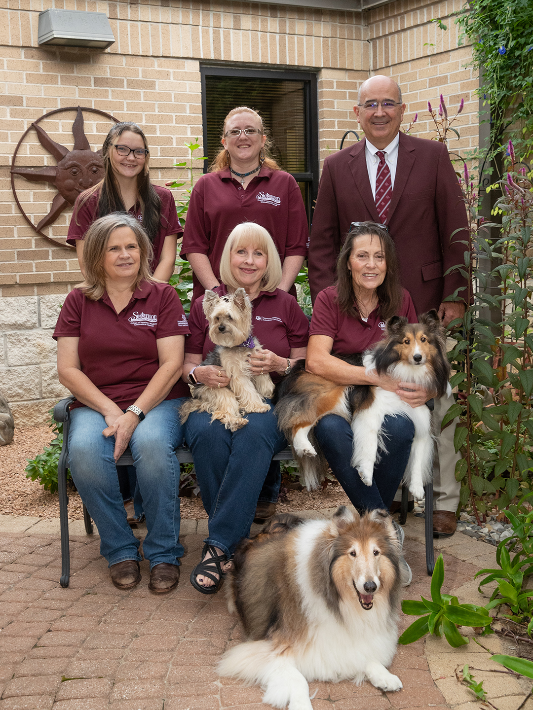
Stevenson Center staff and residents. Back row: Riley Reynolds, Tori Singletary, and Dr. Sam Miller. Middle row: Kimberly Muth, Ellie Greenbaum holding Mitzi, and Janet Broadhead, RVT, holding Cody Mouse. Front row: Miss Reveille IX
Texas A&M University is often recognized as a leader in selfless service. In 1993, the Texas A&M School of Veterinary Medicine & Biomedical Sciences (VMBS) established a center dedicated to selflessly serving pets whose owners can no longer provide their care. The one-of-a-kind center known as the Stevenson Companion Animal Life-Care Center celebrates its 30th anniversary this March.
The Stevenson Center is a one-of-a-kind facility dedicated to serving companion animals whose owners can no longer do so. The center provides a home and embraces pets enrolled in the center as family when they officially become residents at the center. Each resident is cared for by devoted staff; four Doctor of Veterinary Medicine students who live in the center and care for the residents on nights, weekends, and holidays; and veterinarians from one of the highest-ranking schools of veterinary medicine in the U.S.
“Texas A&M is the right place for the Stevenson Center, because of our veterinary hospital and our student residents,” said Dr. O.J. “Bubba” Woytek, assistant vice president of development who helped raise funds to establish the center. “The students who live at the Stevenson Center are veterinarians in training at one of the best schools in the nation, and their teachers and mentors at the teaching hospital offer the Stevenson Center residents veterinary care that’s not available at most veterinary clinics.”
The Stevenson Center was established thanks to the vision of Dr. E. W. “Ned” Ellett, former head of Texas A&M’s Small Animal Teaching Hospital, and monetary donations from the Luse Foundation and Madlin Stevenson, the center’s namesake.
“Mrs. Stevenson loved animals,” Woytek shared. “She had so many animals — several dogs, cats, a horse, a llama — and she loved them all. She was a go-getter who worked as an interior designer until she retired in her 90s.”
In addition to helping fund the Stevenson Center, she also helped decorate the center so that it would feel more homey to residents. Her pets, including her horse and llama, joined the Stevenson Center family when she passed away in 2000. Today, her nephews and nieces-in-law carry on her tradition of supporting the center named in her honor.
Since the center was founded, the facility has undergone two renovations in order to accommodate more residents. A third expansion is underway now and is being overseen by Dr. Sam Miller ‘91, who became the center’s director in the fall of 2022.
“The growth of this program has been pretty significant,” Miller explained. “A big part of our focus is to ensure our facility can accommodate the needs of our residents by providing a safe environment in which they can comfortably interact with the other residents and staff throughout the day, much like their previous homes. The new addition gives us the opportunity to take in more residents and to ensure they enjoy a home-like environment when they join the Stevenson Center family.”
The immense growth the Stevenson Center has experienced is thanks in large part to Dr. Henry L. “Sonny” Presnal ‘57, ‘69, who served as director of the Stevenson Center for 24 years. Under his direction, 122 pets lived out their lives and over 700 pets were enrolled to enter the program.
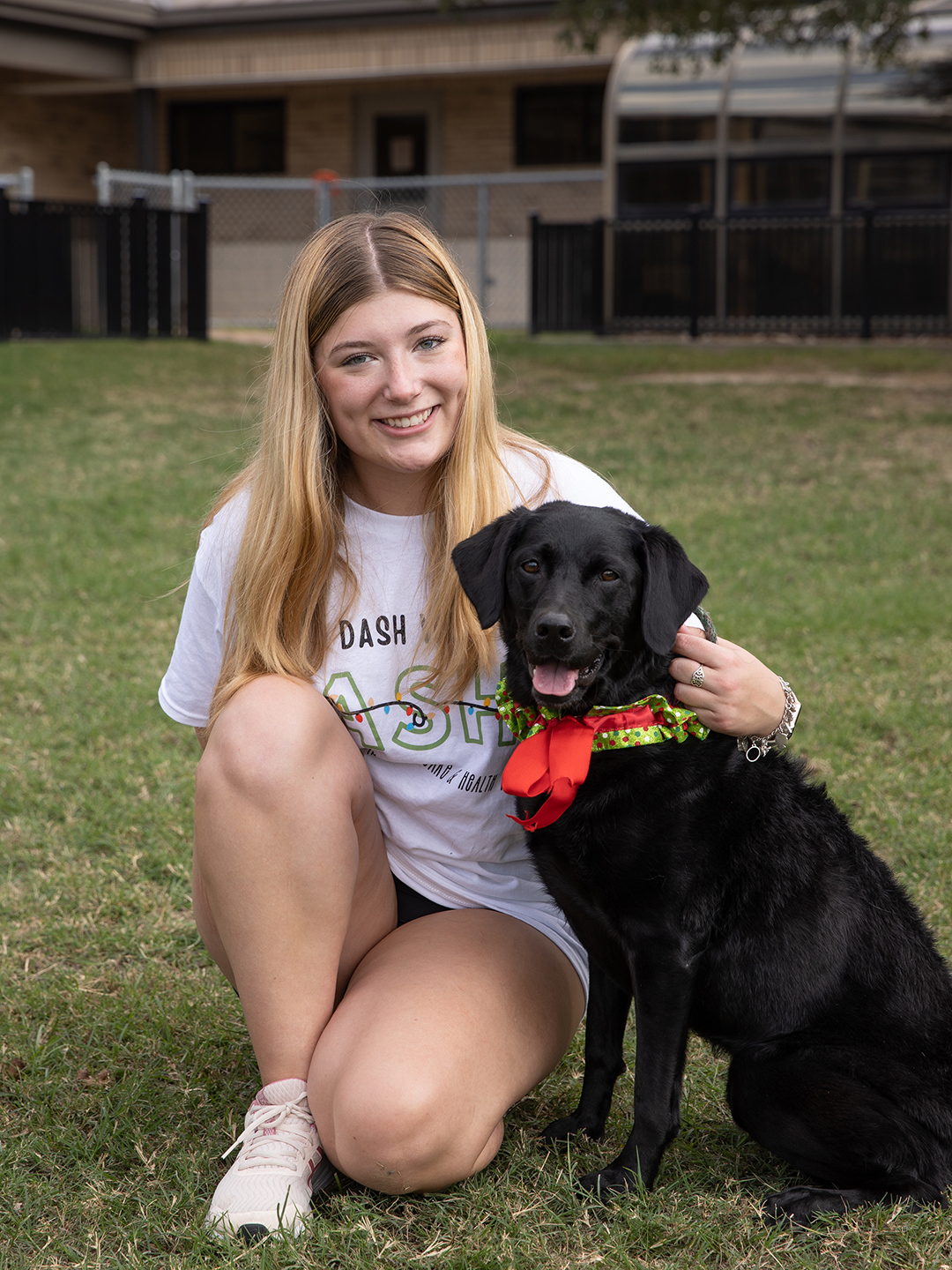
The Stevenson Center provides a home-like environment for veterinary students living at the center and the center’s residents, like April the black Lab.
“Directing the Stevenson Center is a way to serve both animals and people,” Presnal shared. “The thing I cherish most is the relationships I have been able to develop at the Stevenson Center with some of the donors. It’s a relationship that you build over time, and it’s very rewarding.”
Presnal said the Stevenson Center offers the VMBS a unique opportunity to give back to the community.
“It’s important to understand that companion animals are important to the elderly,” he explained. “Humans and animals have a bond that’s special, and the Stevenson Center allows them to live with companion animals without worrying about their care once they can no longer care for their pets themselves.”
Enrolling a pet in the Stevenson Center family requires the establishment of a permanent endowment. The income from this endowment will provide for the care of the pet during its lifetime. Once the pet no longer requires this income the income will be used to support the Stevenson Center and the VMBS.
“A big factor in people being drawn to this program is the fact that we’re affiliated with, located next door to, and supported by the Texas A&M Veterinary Medical Teaching Hospital,” Presnal said, adding that the VMTH provides all medical care to enrolled pets, including wellness and preventative care.
Ellie Greenbaum has served as the associate director of the Stevenson Center since January 1999.
“We have the most excellent staff here, and that’s why it all works,” she shared. “Everybody here loves these animals as much as their original owners love them, and we want nothing but the best for them. It’s an honor to care for these pets, and it makes you feel good that you can offer this service to people and their most beloved family members. It’s like giving us their child and trusting us to take good care of them. We don’t take that responsibility lightly.”
The exceptional care offered to Stevenson Center residents attracts the enrollment of new family members from across the country, with 32 states represented in the center’s current enrollment as well as five from Singapore. In addition to serving companion animals from around the globe, the Stevenson Center is the retirement home for Texas A&M’s mascot, Reveille IX. The center also had the honor of homing Reveille VIII during her retirement as mascot for Texas A&M.
Looking ahead to the Stevenson Center’s next 30 years, Miller said he’d like to see it become a model for future facilities across the nation offering the same service to pets and their families.
“It’s a great opportunity to engage members of the public and offer such a valuable service,” he shared. “We give those pet owners who value the companionship of pets the comfort and confidence in knowing that if their pets outlive them, they’ll not only have a home-like environment, but also a new family to join that is dedicated to providing the highest quality veterinary care available in the country.”
The Stevenson Center will formally celebrate its 30th anniversary with close friends on April 27. Those interested in supporting the Stevenson Center in its next 30 years of service may do so by visiting tx.ag/StevensonDonation.
###
For more information about the Texas A&M College of Veterinary Medicine & Biomedical Sciences, please visit our website at vetmed.tamu.edu or join us on Facebook, Instagram, and Twitter.
Contact Information: Jennifer Gauntt, Director of VMBS Communications, Texas A&M College of Veterinary Medicine & Biomedical Sciences, jgauntt@cvm.tamu.edu,979-862-4216
Kittens and cats, whether they live indoors or outdoors, can be exposed to diseases caused by viruses or bacteria. Vaccinations help protect our feline friends by preventing the spread of these diseases and boosting their body’s defense against potentially fatal illnesses.
Because of this, Dr. Lori Teller, a clinical associate professor at the Texas A&M School of Veterinary Medicine & Biomedical Sciences, encourages owners to vaccinate their kittens and cats against common, yet fatal, diseases sooner rather than later.
“Kittens should begin pediatric visits to the veterinarian when they are 6-8 weeks old, where vaccinations will initially be given as a series until the animal is 16-20 weeks of age,” Teller said. “On the other hand, cats adopted as adults should visit a veterinarian and receive their vaccinations as soon as possible.”
Some common diseases that cats should be protected against include panleukopenia, a viral disease caused by feline parvovirus, and rhinotracheitis, an infection that leads to upper respiratory infections.
“If a cat gets panleukopenia, all of their white blood cells are wiped out, leaving it unable to fight off any other infections; this disease frequently results in death,” Teller said. “Additionally, once a cat has rhinotracheitis, cats will generally become carriers for life and may experience flare-ups of respiratory problems when they become stressed or when their immune system is suppressed.”
Teller explained that there is a combination vaccine that can protect cats against panleukopenia, rhinotracheitis, and calicivirus, a virus that causes upper respiratory infections and ulcers — or sores that develop in the cat’s mouth and on the tongue.”
Additionally, cats who venture outside should be vaccinated against feline leukemia, a highly contagious virus that spreads easily between cats.
“Feline leukemia is most commonly spread through saliva and nasal secretions, so cats that share food and water bowls, groom each other, or bite each other in a fight can spread the virus,” Teller explained. “This virus can lead to a suppressed immune system and leaves a cat susceptible to other infections, as well as cancer, all of which can potentially be fatal.”
There also are vaccines that cats require no matter their lifestyle, such as rabies. Teller pointed out that rabies, which is almost always fatal, can be transmitted to cats by wildlife, including bats, coyotes, raccoons, and skunks.
In addition to vaccinations, Teller suggests that owners protect their cats from diseases spread by mosquitoes, fleas, and ticks.
“Keeping pets on year-round preventives will kill fleas and ticks and prevent the development of heartworm disease and intestinal parasites, which can be infectious to humans,” Teller said.
Finally, a visit to the veterinarian will help determine if your kitten or cat requires any additional vaccinations.
“Your cat’s age, environment, and current health status can help your veterinarian best determine the appropriate vaccinations for your pet to receive,” Teller said. “Vaccinations protect our pets from a variety of diseases that can cause significant illness or death and are always less expensive than the cost to treat illnesses.”
Vaccinations are vital for protecting pets against illnesses and by vaccinating kittens and cats against dangerous diseases, owners can ensure their feline friend lives a healthy and happy life.
Pet Talk is a service of the College of Veterinary Medicine & Biomedical Sciences, Texas A&M University. Stories can be viewed on the web at vetmed.tamu.edu/news/pet-talk. Suggestions for future topics may be directed to vmbs-editor@tamu.edu.