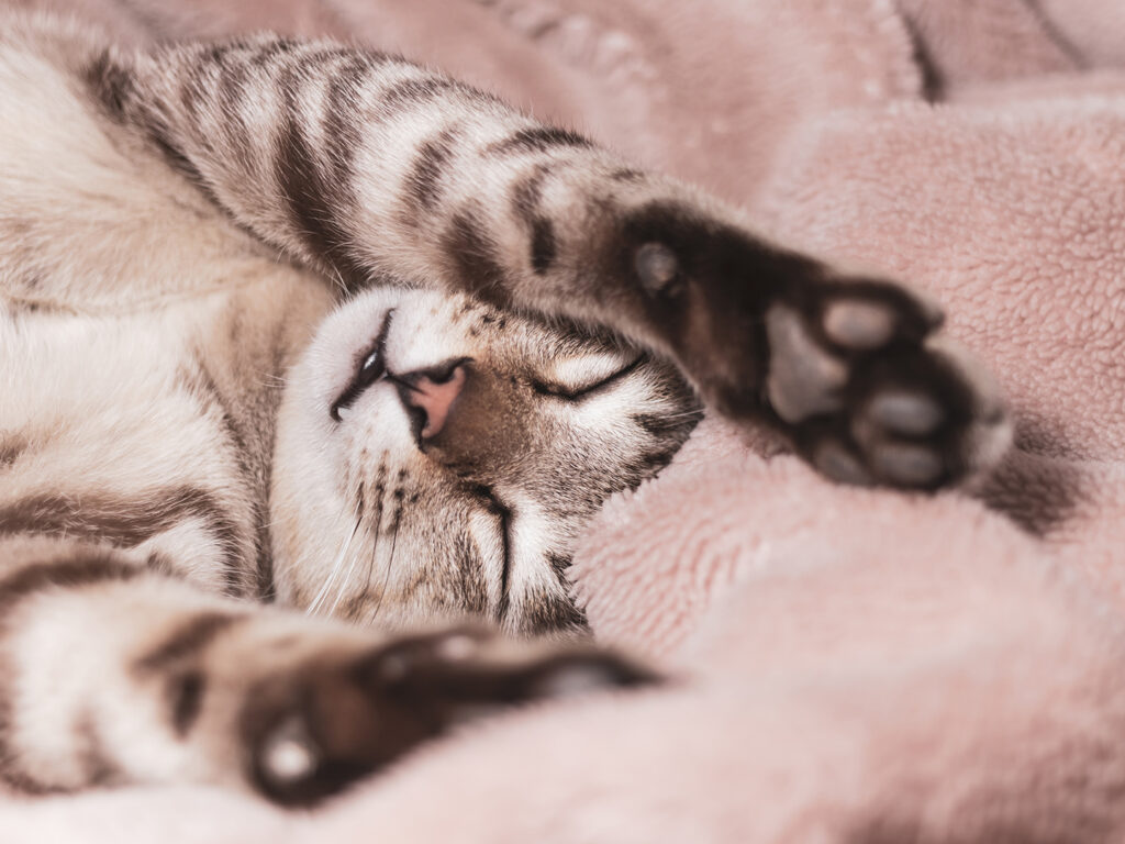
A cat’s paw pads serve a variety of essential functions: they help regulate body temperature through sweat glands; provide traction so that cats can grip surfaces while they climb, jump, or run; and cushion each beautiful, fluid step a cat makes.
Paw pads can also reveal a lot about a cat’s health, as changes in how paw pads look or feel can be a sign that a cat has underlying health issues.
Dr. Christina Gentry, a clinical assistant professor in dermatology at the Texas A&M School of Veterinary Medicine & Biomedical Sciences, provides insight into pillow foot, an uncommon but serious condition that can affect paw pads and prevent cats from engaging in their daily activities.
Identifying, Confirming Pillow Foot
Feline pillow foot, also known as feline plasma cell pododermatitis, is a skin disease that causes a cat’s paw pads to become swollen and painful.
“Pillow foot occurs when high numbers of plasma cells, an immune cell that produces immunoglobulin to help fight infections and regulate the immune system’s response to the infection, infiltrate the tissue of the paw pads, yet the cause for this infiltration is unknown,” Gentry said. “Most cats feel well aside from the discomfort of their paw pads, although a small percentage may act sick with a fever or loss of appetite.”
Owners and veterinarians may notice if a cat has pillow foot by observing changes in the cat’s overall behavior and the appearance of their paws. The big pads of the paw are often affected the most, usually on more than one foot, but occasionally the small toe pads can develop swelling and sores as well.
“During routine interactions or when trimming the cat’s nails, owners may see that the larger pads are soft, squishy, or puffy — sometimes we describe them as feeling like bean bags — and have an open sore,” Gentry said. “In some cases, the first time an owner notices an issue is when the cat is lame, meaning they are less active or not jumping around as much. Other times their veterinarian will notice the swelling and any potentially open sores on the paw pads during physical exams.”
Because there are some conditions with similar symptoms to pillow foot, such as infections or autoimmune diseases, a proper diagnosis for pillow foot is necessary to ensure the cat receives the most effective and appropriate care for their condition.
“Often, pillow foot can be diagnosed during a physical exam if multiple paw pads have a sore or if there is a history of sudden lameness without any known trauma or other skin lesions,” Gentry said. “If the case is more subtle or owners want additional confirmation prior to starting treatment, a tissue biopsy can provide a definitive diagnosis. A fine needle aspirate, which involves poking a needle into the tissue and collecting cells to put on a slide for microscopic analysis, would also support the diagnosis if lots of plasma cells are present.”
What To Expect After Diagnosis
Once diagnosed, the specific treatment approach will depend on the individual cat’s condition and the severity of their symptoms, as more severe cases may require more intensive treatment.
“Oral immunomodulators, which are drug treatments to help regulate the body’s immune system, are the standard treatment and include doxycycline, an antibiotic with immunomodulating properties; and corticosteroids or modified cyclosporine to reduce inflammation,” Gentry explained. “In cases that do not respond to medical management, the pad tissue can be surgically removed.”
Intermittent or long-term treatment also may be necessary, so owners should be sure to follow their veterinarian’s advice closely. This includes attending follow-up appointments to manage their cat’s condition, monitoring their progress, and ensuring their comfort.
“Their veterinarian will slowly taper the immunomodulators and watch for flare ups, but if the condition returns or worsens as the medications are lowered, they may adjust the dose or change medication,” Gentry said. “Some cats require medication for one or two months, while others require an immunomodulator long term, ranging from a few months to a few years.”
With early detection, proper diagnosis, and an appropriate treatment plan for pillow foot, cats can step into a happy, healthy lifestyle, thanks to their owners and veterinary care team.
###
For more information about the Texas A&M College of Veterinary Medicine & Biomedical Sciences, please visit our website at vetmed.tamu.edu or join us on Facebook, Instagram, and X.
Contact Information: Jennifer Gauntt, Director of VMBS Communications, Texas A&M College of Veterinary Medicine & Biomedical Sciences, jgauntt@cvm.tamu.edu,979-862-4216
The NAS is a private, nonprofit institution established under a congressional charter by President Abraham Lincoln in 1863. It is one of three national academies that provide science, engineering and health policy advice to the federal government and other organizations, including the International Science Council.
Elected members are recognized for their achievement in science.
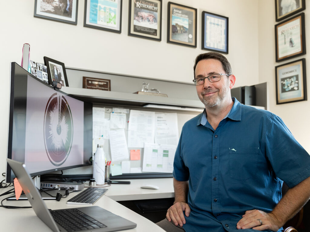
Murphy has received this honor — one of the highest that a scientist can achieve — for his research using comparative genomics to describe and understand evolutionary processes in mammals that result in different species, adaptations and overall biological diversity. His work has helped redefine the mammalian tree of life and transform scientists’ understanding of biology, especially through his research on the domestic cat genome.
His work has even been featured on the covers of highly regarded publications like Science, Nature, Genome Research, the Journal of Heredity, and Genomics.
“Dr. Murphy is one of the most influential scholars in his field worldwide. His seminal scientific achievements have had transformational impacts on the biology of mammals and the health and conservation of cats,” said Dr. John R. August, the Carl B. King Dean of Veterinary Medicine. “We are so proud that Dr. Murphy has received this highest honor for the breakthrough research discoveries that he has made as a faculty member of over 20 years at Texas A&M University. We are also thankful for his service as our assistant dean for the VMBS’ Office of Research and Graduate Studies.”
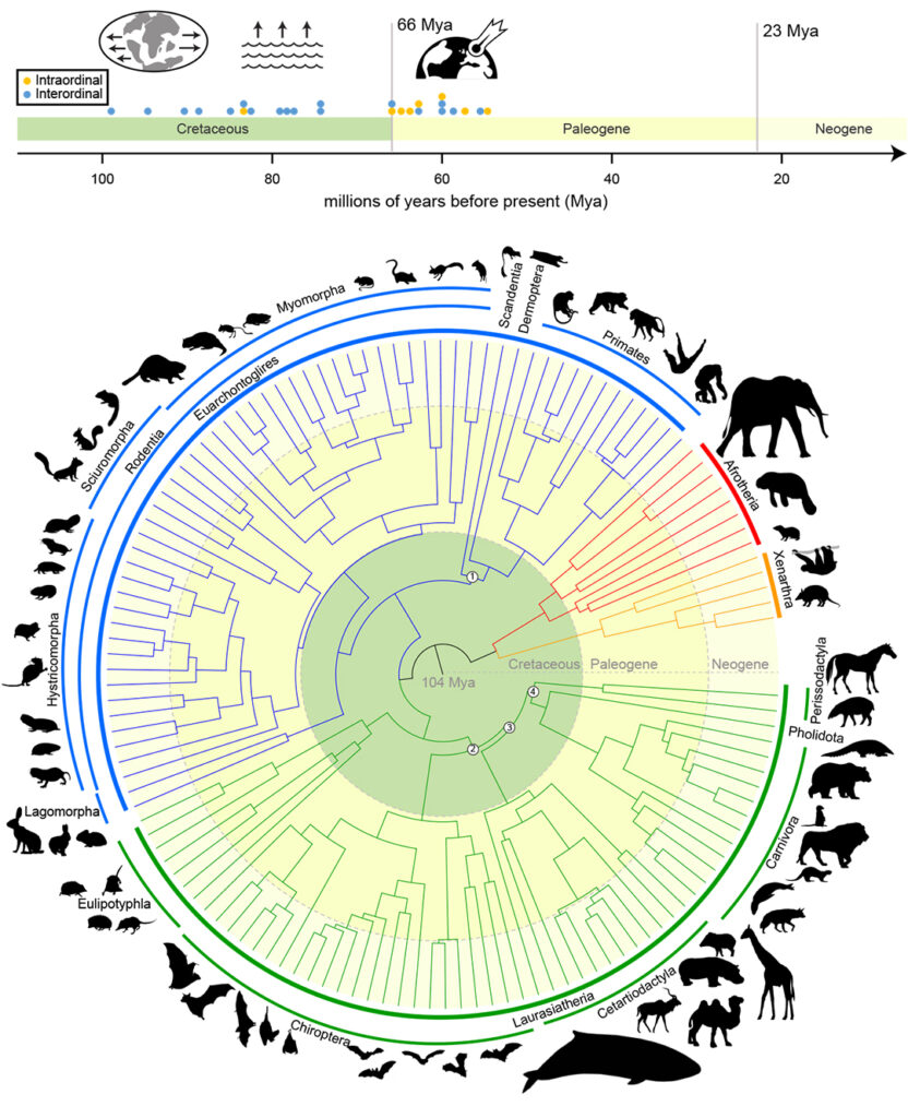
Redefining The Tree Of Life
The “mammalian tree of life” maps out the evolution of mammals over more than 100 million years and is crucial to the goals of the Zoonomia Project.
Some of Murphy’s most transformative work has come from his participation in the Zoonomia Project, an international consortium of scientists who use the largest mammalian genomic dataset in history to answer questions about human evolution as it relates to overall mammal evolution.
In a study published last year, Murphy and his colleagues were able to provide a definitive answer regarding the evolutionary timeline of mammals over the last 100 million years. They found that mammals were already experiencing species diversification even before the mass extinction that wiped out non-avian dinosaurs.
According to the study, mammals saw even more diversification after the extinction because they no longer had to compete with as many dinosaurs for room, resources and stability. This accelerated rate of diversification is why there are so many different mammal lineages, including carnivores, primates and hoofed animals.
The ability of scientists to compare the human genome with so many mammal genomes using the Zoonomia Project’s data has led to important discoveries impacting health and medical treatments for both animals and human beings.
Unveiling The Secrets Of Cat DNA
Murphy is also regarded as an expert in cat evolution and genomics, with his work providing the foundation for medical treatment development and conservation efforts that promote feline health and well-being. Some of his recent research has helped scientists better understand what makes different cat species unique, including how they have adapted to their environments.
For example, Murphy’s research into cat olfactory genes — the parts of cat DNA that govern their ability to smell — has revealed important differences between lions and tigers. Tigers, as it turns out, have a larger repertoire of genes that enhance their ability to smell and detect pheromones.
This is likely because tigers are solitary animals that need to be able to detect prey and potential mates across large territories, while lions typically live in much closer proximity to other lions.
While discoveries like this one help scientists better understand species behaviors, they also contribute directly to conservation efforts. For example, understanding how different populations of tigers have evolved to live in different environments — from hot, humid jungles to snowy tundras — helps make it clear to policymakers that these populations are not interchangeable.
A Distinguished Career
Murphy received his Ph.D. in biological sciences in 1997 from The University of Tulsa before becoming a postdoctoral researcher — and then senior scientist — at the Laboratory of Genomic Diversity at the National Institutes of Health’s National Cancer Institute.
After joining Texas A&M in 2004, Murphy was named a Presidential Impact Fellow in 2017 and a University Professor in 2020. Over the course of his career, he has published more than 130 articles in peer-reviewed journals and currently serves as the editor in chief of the Journal of Heredity.
Only 14 other living faculty members at Texas A&M are NAS members; the only other member from the VMBS is Dr. Leif Andersson, also a VIBS professor.
“We are very proud of Dr. Murphy and are excited to see how he continues to change the field of comparative genomics,” said Dr. Michael Criscitiello, a professor in the VMBS’ Department of Veterinary Pathobiology and the associate dean for the VMBS’ Office of Research & Graduate Studies. “It’s very fitting that he has been given this honor, since he is the James E. Womack University Professor of genetics, and the late Dr. Womack was also a member of the NAS.”
“Bill’s work has provided fundamental advancements in phylogenetics and genomics,” said Dr. Alex C. Keene, professor and head of the Texas A&M Department of Biology, where Murphy holds a courtesy joint appointment. “He has elevated the stature of research at Texas A&M and, more broadly, as the chief editor of the Journal of Heredityfor the last seven years. He has always taken the time to support junior faculty and students across the campus in a way that has advanced the careers of dozens of younger scientists. This is really so exciting for Texas A&M, and we are all so happy for Bill!”
###
For more information about the Texas A&M College of Veterinary Medicine & Biomedical Sciences, please visit our website at vetmed.tamu.edu or join us on Facebook, Instagram, and X.
Contact Information: Jennifer Gauntt, Director of VMBS Communications, Texas A&M College of Veterinary Medicine & Biomedical Sciences, jgauntt@cvm.tamu.edu,979-862-4216
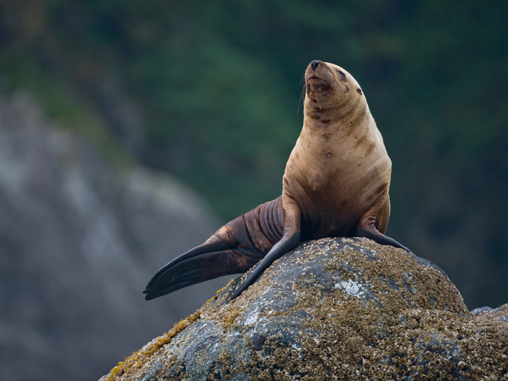
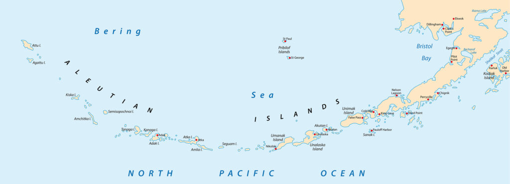
Although Steller sea lions were added to the endangered species list in the 1990s, their populations began recovering in some parts of the Aleutian Islands in the early 2000s.
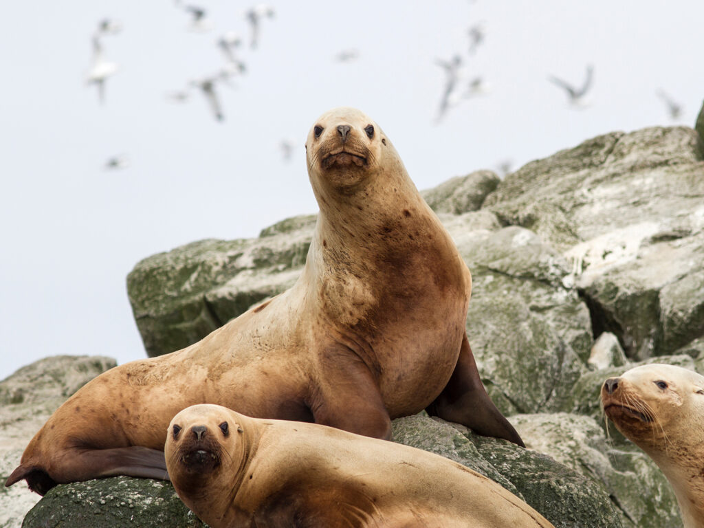
A team of researchers from Texas A&M University and beyond has made a surprising discovery about rising mercury levels in Steller sea lion pups that may have detrimental effects on the endangered species.
The team’s decade-long effort to study mercury in Steller sea lions in the Aleutian Islands — the strip of islands stretching between Russia and Alaska and separating the Bering Sea and Pacific Ocean — has revealed that the number of pups born with potentially dangerous levels of mercury in their blood and fur increased by more than 50% from 2011 to 2018 before leveling off in 2019.
Mercury — a “heavy metal,” non-essential element — can be toxic to some animals, including humans, at high concentrations. Various forms of mercury can be introduced into the environment via emissions from human activities; it can also be introduced naturally through seismic activity (like volcanoes) and melting permafrost, making it harder to pinpoint the cause of it increasing in the food web.
Before joining the Texas A&M faculty as a professor of veterinary toxicology in the School of Veterinary Medicine and Biomedical Sciences (VMBS), Dr. Todd O’Hara was a toxicologist in Alaska who conducted studies on whether mercury levels were safe in the ocean and the food web.
“While our findings haven’t indicated an immediate threat to humans via what we eat from this part of the ocean, they have raised questions about how mercury and other elements may continue to change in some of the fish we eat now as rising ocean temperature trends continue,” O’Hara said
The levels of mercury found in the pups in a largely remote area led the team to extend its research efforts in search of the cause and to examine changes in other elements in Steller sea lion populations.
The team is trying to determine whether there is a correlation between higher mercury levels in sea lion pups and population decline.
‘Heavy Metals’ And Health
Steller sea lions were added to the endangered species list in the 1990s, but their populations began recovering in some parts of the Aleutian Islands in the early 2000s, with the eastern distinct population being delisted in 2013. To discover why some groups remained in decline, researchers teamed up with the National Oceanic and Atmospheric Administration (NOAA) and commercial fisheries via Ocean Peace Inc. to gather information about the different populations from rookeries where pups and their prey are studied.
“When we started this research in Southeast Alaska, we found very few pups with alarming levels of mercury,” said Dr. Lorrie Rea, a research professor at the University of Alaska Fairbanks (UAF) who plays a significant role in the team’s Steller sea lion research program. “Once we got to the Western Aleutian Islands, we found pups with mercury concentrations that were three to four times higher than the highest we saw in other regions. As we moved toward Russia along the Aleutian Island chain, the concentrations went down again, so it’s almost like a bell-shaped curve.”
The researchers had discovered a region of higher mercury concentrations in Steller sea lion pups along this island chain.
The researchers also found that among the populations in the different regions of the Aleutian Islands, those with higher mercury levels overlapped with the populations still struggling to recover their numbers. This led the team to investigate mercury increases as a possible contributor to the sea lion’s lack of population recovery.
The team found a relationship between Steller sea lion pups with high mercury levels and certain kinds of fish in their mothers’ diets.
O’Hara said the major concern is the transfer of mercury to the fetus, which is vulnerable to adverse effects.
“Mercury is known to negatively impact immune health in humans, and one of Todd’s graduate students, Stephanie Kennedy, determined that it has a similar effect in animals like Steller sea lions,” Rea said.
Mercury levels can also affect the body’s ability to resolve oxidative stress, which harms overall health by keeping antioxidant levels relatively low and causing tissue breakdown over time. Thus, the team decided to include many essential elements, such as selenium, in the research effort.
“Oxidative stress is especially important for diving mammals such as sea lions because they go through long breath holds when they go to forage, or hunt, for fish,” Rea said. “The longer breath holds use up a lot of oxygen, so having this ability to counter the oxidative stress is important for their everyday foraging.”
O’Hara said many selenium-dependent processes are known to be involved in this protection and also plays a role in protecting from mercury toxicosis, or poisoning.
The researchers discovered a region of higher mercury concentrations in Steller sea lion pups along the Aleutian Island chain.
Metal Detectors
After discovering differences in mercury levels between Steller sea lion populations at different points along the Aleutian Islands, the team set out to determine if other elements also varied in pups.
The team worked with Dr. Daniela “Hanny” Alejandra Murillo Cisneros, a post-doctoral researcher at Centro de Investigaciones Biológicas del Noroeste located in La Paz, the capital city of Baja California Sur, Mexico.
Cisneros is lead author on a recently submitted manuscript reporting on these elements in Steller sea lions. She uncovered differences in numerous elements in hair and blood of Steller sea lion pups among populations across the regions of the Aleutian Islands. In hair, all 12 elements measured were different by region, except zinc, based on a distinct oceanographic feature, Amchitka Pass.
Her discovery showed that the geographic locations of the populations are playing a significant role in the Steller sea lions’ exposure to both nonessential elements, which are potentially toxic, and essential elements, which are required for good health. Thus, investigators need to consider deficiencies of essential elements and not just those that may be harmful or toxic.
“You are what you eat, and this research reflects that,” Cisneros said. “What we found is that there are significant chemical composition differences in the food sources available at each of these regions, and those differences are being reflected in Steller sea lion populations. We are taking a very close look at prey now, and some of the prey species are important food for humans.”
Cisneros’ elemental discoveries also provide evidence that the oceanic Amchitka Pass — located near the center of the Aleutian Islands where the previously mentioned “bell-shaped curve” spikes in Steller sea lion pup mercury levels — is functioning as a barrier beneath the ocean’s surface that seems to drive the varying chemical composition of Steller sea lions east and west of the pass.
“We can’t visually see some of the barriers or divisions in the ocean because they’re so deep and they’re so complicated,” O’Hara said. “Based on our studies in fish and Steller sea lions, we’re showing that this simple pass has divided the ocean biologically, physically and chemically.”
Implications For Human Health
The geographic locations of Steller sea lion populations are playing a significant role in their exposure to both nonessential elements, which are potentially toxic, and essential elements, which are required for good health.
Fish in grocery stores around the world come from the same waters that Steller sea lions forage in, making it important for humans to pay close attention to mercury and other elements in the Steller sea lion diet. The team considers the sea lion as an environmental sentinel.
Thankfully, the fishing industry is helping research teams study mercury levels by donating fish to academic research and financial support for the trace elements analysis conducted at the VMBS. So far, commercial fisheries have donated about 1,500 fish from the Aleutian Islands region.
“Everyone working on finding these answers, from the researcher to NOAA to the fisherman who donate fish, we all want science-based decision making,” O’Hara said.
Because of the support from the fishing industry, the researchers have been able to test the donated fish for diet markers, mercury and trace elements such as selenium.
“The good thing for human health is that of the 1,500 fish, we only found about 13 that had mercury concentrations that were above the levels advised for human consumption, and most of the 13 were yellow Irish lord, which is a spiny fish not typically included in fish sold to humans,” Rea said.
Fishing For Answers
The researchers emphasize that they’re not jumping to conclusions about what’s causing the rising mercury levels or the stalled sea lion population recovery on some islands.
“It’s important to note that these findings so far prove correlation, or that these things happened at the same time, not causation, which would mean one event caused the other,” O’Hara said.
As the research team continues its work in the Aleutian Islands, O’Hara, Rea, and Cisneros said they’re thankful for the collaborative nature of their work as toxicologists.
“Discoveries like this and the continued hunt for answers really takes a village,” Rea said. “This research brings academics from around the world together who contribute multiple perspectives as well as support from various industries and agencies with a vested interest in what we’re finding.”
The teams learn from each other across the border to determine what are the best approaches to understanding trace elements in the Pacific Ocean and changes in the ocean temperature and pH.
O’Hara said this work highlights the fact that how people impact the ocean has many potential effects that are not intuitively obvious, and changes in mercury levels in ocean life may be driven by many factors.
“We find answers in individual projects, but they come together like puzzle pieces to build a bigger picture,” Rea said. “We won’t fully understand what’s happening with Steller sea lions until we understand what’s happening to their environment, so there’s a lot more work to be done with other agencies and continuing our academic and industry collaboration is key to our success.”
Other researchers involved with discovering the regional differences include those from the Trace Elements Research Laboratory (TERL) in the Department of Veterinary Integrative Biosciences (VIBS). The related and relevant work in Mexico (led by Cisneros) includes published mercury and selenium concentrations findings in free-ranging California sea lions in the Gulf of California, Mexico. Contributors to the international research in Mexico and Alaska include Dr. Robert Taylor, TERL director; Dr. Jill Hiney from VIBS; Dr. Carlos A. Rosado Berrios from VIBS; Dr. Ben Barst from UAF; and graduate student Michelle Trifari from UAF; among many others.
###
For more information about the Texas A&M College of Veterinary Medicine & Biomedical Sciences, please visit our website at vetmed.tamu.edu or join us on Facebook, Instagram, and X.
Contact Information: Jennifer Gauntt, Director of VMBS Communications, Texas A&M College of Veterinary Medicine & Biomedical Sciences, jgauntt@cvm.tamu.edu,979-862-4216
Kayleigh Shumaker at the 2023 National Veterinary Scholars Symposium
Second-year veterinary student Kayleigh Shumaker has received two prestigious awards for her research on Down syndrome at the Texas A&M School of Veterinary Medicine & Biomedical Sciences (VMBS).
Shumaker was one of five students nationwide, and the first ever from Texas A&M, selected for the American Veterinary Medical Foundation’s 2nd Opportunity Summer Research Stipend, as well as the sole recipient of the Boehringer Ingelheim (BI) Veterinary Student Award.
The 2nd Opportunity Summer Research Stipend of $6,000 will support Shumaker’s second summer participating in the VMBS’ Veterinary Medical Scientist Research Training Program (VMSRTP), which allows veterinary students to conduct full-time research during a 13-week period in the summer under the advice and direction of a faculty mentor.
As the BI Veterinary Student Award recipient, Shumaker will receive a $1,500 award, travel to Minnesota, and attend the 2024 National Veterinary Scholars Symposium in August to give a plenary oral presentation on her VMSRTP research project.
“My project was looking at altered angiogenesis, or blood vessel formation, as a potential mechanism behind impaired fracture healing in Down syndrome,” Shumaker said. “People with Down syndrome are predisposed to low bone mineral density and are at increased risk of fracture nonunion, or fractures that don’t heal. This is a huge problem because fractures that don’t heal have a real impact on quality of life.
“What we found suggests that changes in blood supply and blood vessel formation are partially responsible for these fractures not healing, which is exciting to uncover and prompts more questions, as research always does,” she said.
Shumaker will continue her work in Suva’s lab during her second summer in the VMSRTP, working with Ph.D. student Catrina Silveira, DVM, to continue studying angiogenesis in fracture healing and contribute to other similar projects.
“We are, indeed, proud of Kayleigh as an outstanding representative of the VMSRTP as well as of the Texas A&M Doctor of Veterinary Medicine program,” said Dr. Dana Gaddy, a VMBS professor in the Department of Veterinary Integrative Biosciences and VMSRTP director. “We are delighted to champion Kayleigh’s success and welcome her back to VMSRTP as a senior scholar as well as to Dr. Suva’s lab to work with Dr. Silveira to understand compromises in fracture healing in Down syndrome.”
New Career Goals
Shumaker first developed an interest in research because of her desire to pursue a career in small animal surgery.
“I had zero research experience coming into vet school, but I knew I had an interest in small animal surgery and part of pursuing an internship and residency is getting research experience,” she said. “So, I applied to the VMSRTP, which ended up being a highly impactful experience with great mentorship that encouraged me to pursue research for another summer.”
Her ultimate career goal is to work as a veterinary surgeon in academia so that she can continue to pursue research while also teaching and caring for patients.
“These awards are a testament to trying new things and the excellent mentorship I received through the VMSRTP,” Shumaker said. “If it weren’t for the support from Dr. Larry Suva, Dr. Dana Gaddy, Dr. Catrina Silveira, and others in the lab, I would not have been able to complete my project and certainly would not have received these awards.”
###
For more information about the Texas A&M College of Veterinary Medicine & Biomedical Sciences, please visit our website at vetmed.tamu.edu or join us on Facebook, Instagram, and X.
Contact Information: Jennifer Gauntt, Director of VMBS Communications, Texas A&M College of Veterinary Medicine & Biomedical Sciences, jgauntt@cvm.tamu.edu,979-862-4216
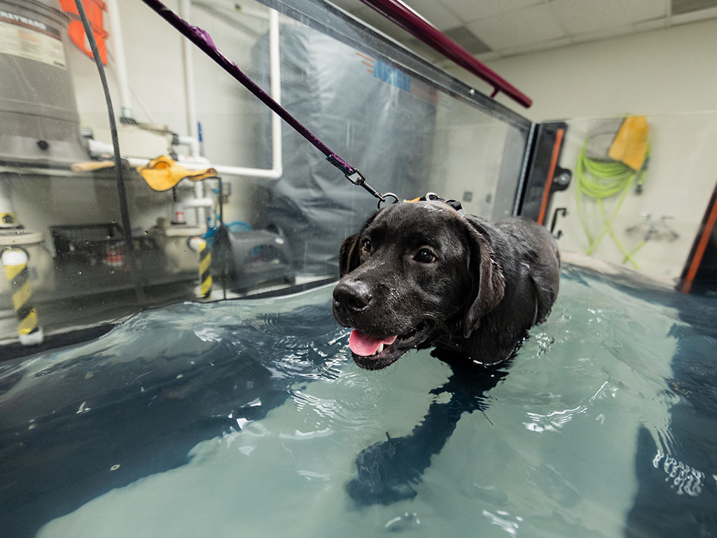
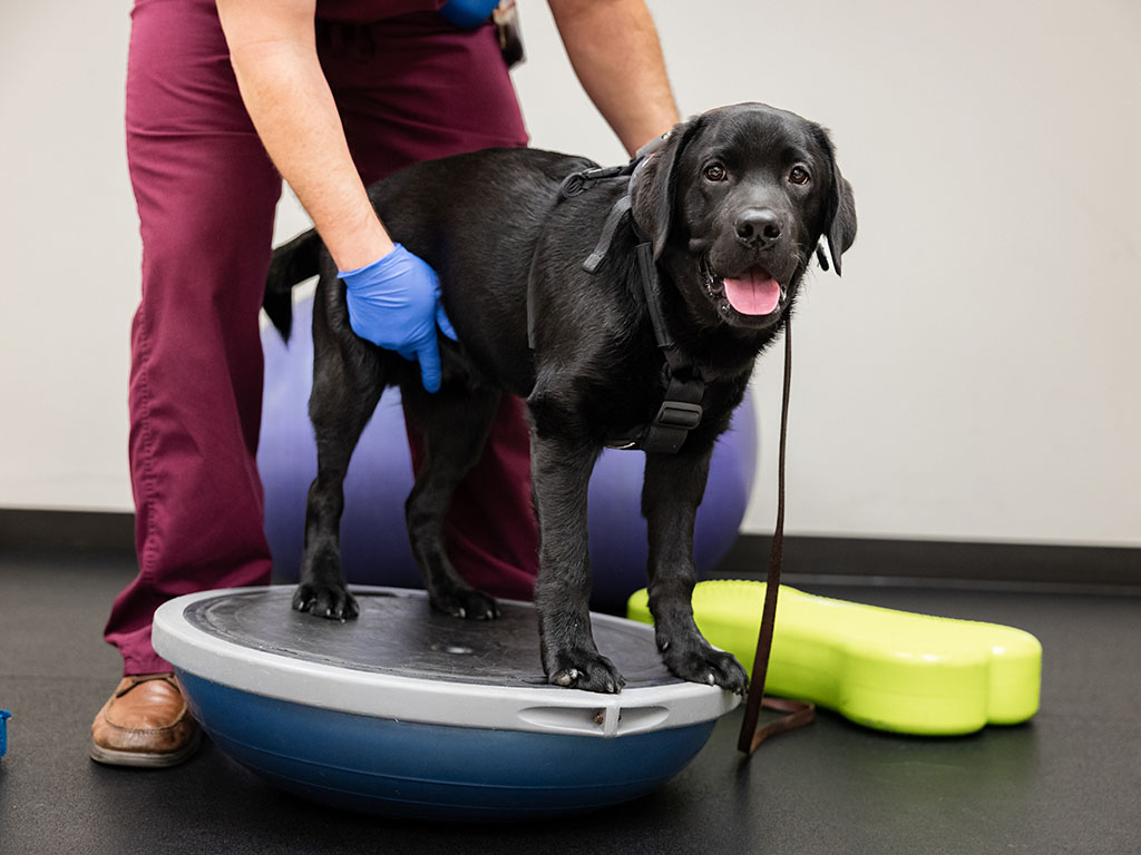
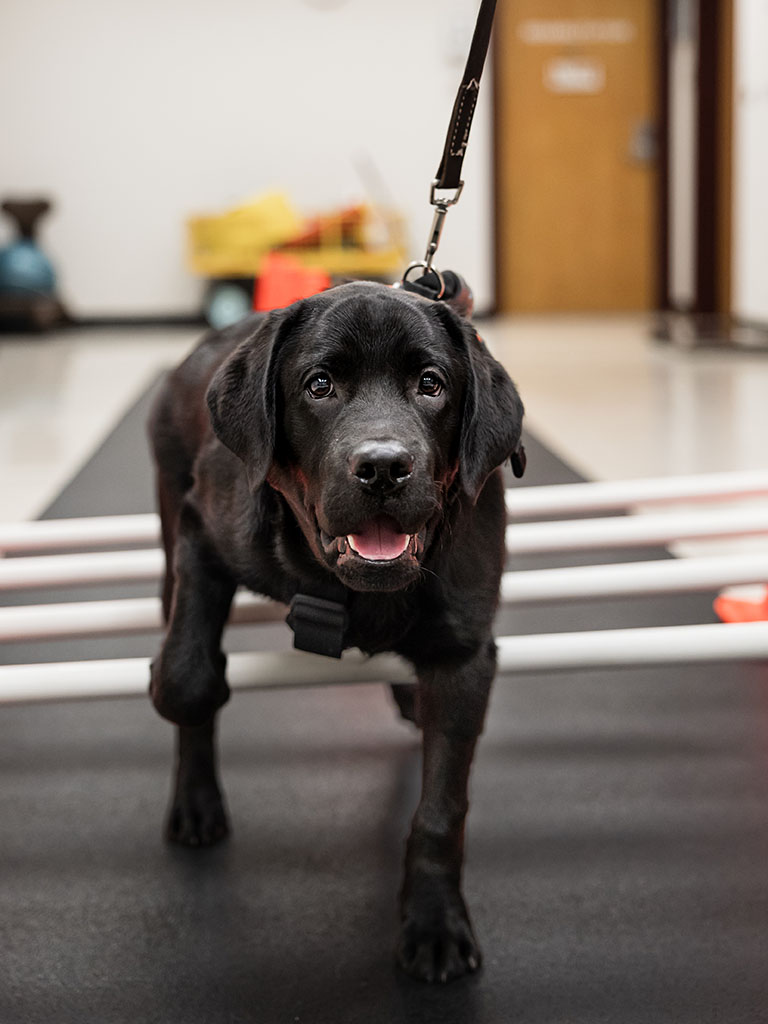
Parlay exercises on the SATH’s water treadmill Photo by Jason Nitsch ‘14, Texas A&M School of Veterinary Medicine & Biomedical Sciences
Parlay, a black Labrador retriever, was named after a gambling term that refers to a series of bets.
When the little lab was diagnosed with tetanus — a serious disease of the nervous system caused by a toxin-producing bacteria — his name took on a new meaning as his veterinary team at the Texas A&M Small Animal Teaching Hospital (SATH) worked to help him beat the odds and survive his condition.
While humans are highly susceptible to the toxin-producing bacteria and are vaccinated regularly for tetanus, there is no tetanus vaccine available for dogs because they are relatively resistant to the disease, according to the National Institutes of Health. However, when dogs contract tetanus, they can experience many of the same symptoms, including lockjaw, stiff muscles that make it impossible for them to bend their legs, muscle spasms in the face, and uncontrollable tightening in the diaphragm, making it difficult to breathe.
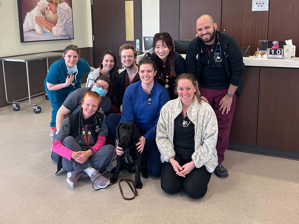
After a nearly two-week stay at the SATH with the Emergency & Critical Care Service and months of physical therapy with the Sports Medicine and Rehabilitation Service, Parlay is back in good health.
A Special Connection
Parlay, an energetic puppy with a loving and curious personality, was the pick of the litter when he was born into a show dog family.
“He’s such a great puppy, and he’s always been like that,” said Wendy Knox, Parlay’s owner. “I stayed with him and the rest of his litter 24/7 for the first four weeks of their lives. That’s something I do with every litter. Because of that, I know all the puppies in a litter pretty well, but Parlay is really special.”
Knox was first introduced to the world of show dogs through her parents, who picked up the new hobby once she and her sister had left home for college. She eventually took over showing the dogs for her parents, but in 2012, the family took a break when Knox’s mom was diagnosed with cancer.
Parlay performs a rehabilitation exercise at the SATH Photo by Jason Nitsch ‘14, Texas A&M School of Veterinary Medicine & Biomedical Sciences
“She got pancreatic cancer,” Knox shared. “She lived for almost 19 months and then lost her battle. We miss her terribly bad. The dogs are a connection to my mom now and something I still enjoy doing with my dad and now my husband, too.”
As part of the Knox family, Parlay has a well-rounded routine. Knox and her husband moved into a home on her parents’ 7 acres near Austin, so Parlay has plenty of space to train and gets to see Knox’s father regularly. There, Parlay enjoys long walks, regular swims in a pond, playful training sessions, and lots of love from the family.
“I spend all my waking time, outside of my job as a program director for the Texas Department of Transportation, taking care of my dogs and walking them on our property, swimming with them, and training them,” Knox said. “Parlay really enjoys playing outside and swimming in the pond with his cousin, Kinslee. He’s just the best dog and is very special to me.”
High Stakes Disease
On what started as a normal Saturday in December, Parlay enjoyed playtime outside with Kinslee and then came in for a nap, which was when Knox noticed a small, swollen bump on the left side of Parlay’s face.
“I examined it and thought it might be a bee sting,” Knox shared. “He was acting healthy and normal, so I decided to keep an eye on him for the rest of the day in the house and give him some Benadryl.”
Parlay ended the day seemingly healthy, with no new developments from the bump on his face. The next day, however, Parlay appeared to have some stiffness, so Knox made an appointment with their local veterinarian for Monday morning.
“Sunday evening, we decided he was probably experiencing a scorpion sting instead of a bee sting, but overall, he still seemed pretty healthy,” Knox said. “That was not the case on Monday morning. He was having trouble getting off his doggy bed and when he did, he was much stiffer. Worst of all, it seemed like he was having trouble breathing.”
Knox wasted no time getting Parlay to his veterinarian. When the veterinary team heard he was having trouble breathing, they, too, sprang into action and saw Parlay right away.
“They took Parlay to the back of the clinic, and when they came to see me about 15 minutes later, they said, ‘Your puppy has tetanus,’” Knox shared. “I looked at them blankly and kept asking, ‘What’s tetanus in a dog?’”
Parlay with members of the veterinary team that saved his life after being diagnosed with tetanus.
Tetanus — also called lockjaw because it causes severe tightness in the jaw and neck, causing the jaw to “lock” — is caused by the bacteria Clostridium tetani, which is found everywhere in the natural environment, including soil, dust, and manure, according to the Centers for Disease Control and Prevention.
The veterinary team explained that the severity of effects from the toxin on the nervous system make the swift administration of antitoxin key to patient survival, while treatment of any conditions or additional ailments that result from the infection are important for assuring quality of life.
Knowing Parlay would need advanced emergency veterinary care around the clock, they informed Knox that she needed to get Parlay to the Texas A&M School of Veterinary Medicine & Biomedical Sciences.
Betting On Parlay
The Bastrop-based veterinary team immediately began calling other clinics to find antitoxin that could be administered to Parlay.
“Since no other emergency veterinarian clinic in the Austin area had the antitoxin, the Bastrop Veterinary Hospital provided me the antitoxin they had on hand for horses” Knox shared. “They packaged me up with two bottles and sent me to Texas A&M. I can still remember them saying, ‘You need to drive and get there as safely as you can but also as quickly as you can. If you don’t, he’s going to die.’”
At the SATH, Parlay and Knox were greeted by Dr. Cody Riffe, a resident clinician on the Emergency & Critical Care Service, who assured Knox they would do everything they could to save Parlay.
“We just didn’t want to give up on him,” Knox said. “As a family, we knew we’d do whatever we could to save him and we knew he was in good hands at Texas A&M.”
As Parlay and his veterinary team continued to combat the bacteria, Parlay’s body experienced severe symptoms, which led to additional ailments, a common challenge with tetanus patients.
Parlay performs a rehabilitation exercise at the SATH Parlay performs a rehabilitation exercise at the SATH Photo by Jason Nitsch ‘14, Texas A&M School of Veterinary Medicine & Biomedical Sciences
“Parlay had difficulty eating and drinking due to the lockjaw; limb weakness as a result of the severe tightening of the muscles that made it so that he was unable to walk or support his own weight; and aspiration pneumonia, likely a result of the toxin attacking the nerves in his throat and esophagus which allowed saliva or food to enter his airway,” Riffe shared. “Our team worked around the clock to make sure Parlay had the care he needed to combat all his ailments.
“He was here for almost two weeks and during that time, his muscles became locked in an extended position,” Riffe said. “Muscle strength will quickly decline without the constant activity of bearing weight and moving through space, so we referred him to the SATH’s Sports Medicine & Rehabilitation Service to help him strengthen his muscles and live a more comfortable, activity-filled life.”
While Parlay was much improved, he still needed assistance standing and walking when it came time to be discharged from the SATH, so he was sent home with a full body harness that supported his weight.
“We got home at about 1 p.m. on Thursday, and on Friday, he started trying to walk on his own with his weight supported by the special harness,” Knox said. “By Friday night, he was able to walk again, and it was the coolest thing to see him doing so much better.”
Parlaying Veterinary Care Into Health
Now, almost four months after his initial diagnosis, Parlay is easing back into his show dog routine so that he’ll be ready for his first show season.
“He enjoys playing outside again, taking his naps, and getting all the love and attention,” Knox said. “We incorporated some of the exercises we learned in physical therapy into his walks around the property, like having him walk backwards to help rebuild muscles in his legs. As the weather warms again, we’re also going to have him start swimming in our pool for hydrotherapy.”
Knox said she’s incredibly thankful for the care Parlay received at the SATH.
“Every single person I’ve encountered at the Small Animal Teaching Hospital — it doesn’t matter whether they’re in the pharmacy or billing or serving as a technician or clinician — everybody was so nice, professional, and considerate,” Knox said. “I have nothing but good things to say, and that’s why I’d recommend anyone in a similar situation bring their animals to Texas A&M.”
###
For more information about the Texas A&M College of Veterinary Medicine & Biomedical Sciences, please visit our website at vetmed.tamu.edu or join us on Facebook, Instagram, and X.
Contact Information: Jennifer Gauntt, Director of VMBS Communications, Texas A&M College of Veterinary Medicine & Biomedical Sciences, jgauntt@cvm.tamu.edu,979-862-4216
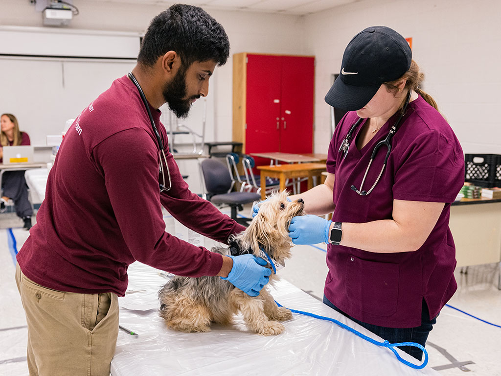
DVM students Mohan Iyengar and Cheryl Armstrong examine a patient at Operation Border Health Preparedness. Photo by Jason Nitsch ‘14, Texas A&M School of Veterinary Medicine & Biomedical Sciences
The Texas A&M Veterinary Emergency Team (VET) was part of a collaborative effort by Texas A&M University to support border health that has received the 2024 Excellence in Interprofessional Education Collaboration Award.
Awarded by the United States Public Health Service Commissioned Officers Foundation for the Advancement of Public Health and the Interprofessional Education Collaborative, the award is presented to a team of health students and faculty whose interdisciplinary work has significantly impacted the community they serve.
“The Texas A&M Veterinary Emergency Team sets the global standard for veterinary disaster response,” said Dr. John R. August, the Carl B. King Dean of Veterinary Medicine at the VMBS. “Their efforts at Operation Border Health Preparedness, in collaboration with their peers from other Texas A&M University entities, highlight the selfless service inherent to both animal and human medicine.”
An Award Worthy Effort
Three faculty members at the Texas A&M University Health Science Center — Drs. Asim Abu-Baker, LeRoy A. Marklund, and Kelly Sopchak — applied for the award on behalf of all Texas A&M affiliated OBHP participants.
“It’s an honor to serve with colleagues from across campus to help our fellow Texans,” said Dr. Deb Zoran, director of the VET.
Abu-Baker, associate dean for clinical and professional affairs at the Irma Lerma Rangel School of Pharmacy, served as principal investigator for the application. Marklund, clinical assistant professor at the School of Nursing, and Sopchak, a psychologist in the Department of Psychiatry and manager of the Texas Child Health Access Through Telemedicine (TCHATT) program at the School of Medicine, represent the 2023 OBHP planning committee.
The interprofessional team that served on the committee also includes Zoran; Dr. Karen Beathard, instructional associate professor in the Department of Nutrition; Dr. Stephen “Eric” Grayson, assistant professor of pharmacy practice in the School of Pharmacy; Dr. Krystal Flores, instructional assistant professor in the School of Public Health; and Dr. Garett Sansom, research assistant professor in the School of Public Health.
Operation Border Health Preparedness
OBHP is an annual, week-long exercise hosted by the Texas Department of State Health Services that provides emergency response teams the opportunity to test their readiness for the next major disaster while also providing annual medical and veterinary care to communities that would otherwise go without.
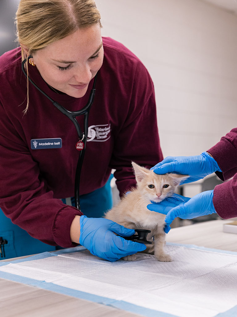
DVM student Madeline Iselt conducts an exam at OBHP. Photo by Jason Nitsch ‘14, Texas A&M School of Veterinary Medicine & Biomedical Sciences
The most recent operation took place in five cities across the Rio Grande Valley — Brownsville, Laredo, Raymondville, Rio Grande City and San Juan — and provided numerous services for more than 6,000 patients, including free physicals, screenings, dental care, immunizations, vision exams, free eyeglasses, and veterinary services for cats and dogs. In this medically underserved region, this event is many residents’ sole opportunity to receive care, and many line up for hours — even overnight — to secure their spot.
The VET’s role in the annual exercise includes providing veterinary care in the South Texas community of Raymondville, which doesn’t have a veterinarian. The team convoys several hundred miles to South Texas, spends a couple of days training veterinary students and volunteers while also establishing a base of operations, and then provides annual checkups and immunizations to the resident companion animals from the community for the next five days. These wellness checks, deworming treatments, and vaccinations protect both pets and the people who love them from zoonotic diseases, which are illnesses that can be spread between humans and animals, such as rabies.
“OBHP approaches public health holistically,” Zoran said. “While we provide care to cats and dogs and help protect both them and their families from illness, our colleagues in human medicine provide people in the community essential health care. Together, we’re serving Texans in need while also ensuring our ability to respond to the next major disaster.”
In 2023, the VET completed a record 1,022 veterinary visits, which almost doubled the number of patients seen in 2022 and was significantly higher than the team’s caseload in 2021, the first year the VET participated in OBHP.
Hands-On Learning
The volume of patients seen in such a short amount of time provides a huge amount of experience for the Doctor of Veterinary Medicine students who accompany the team.
“Students who serve with us at OBHP get to participate in a full-scale disaster response exercise with hands-on participation in establishing a base of operations and providing basic veterinary care,” Zoran shared.
The experience is invaluable according to Daniel Martinez, a student who traveled to OBHP with the VET in 2023.
“I built relationships with my classmates, teachers and other personnel that demonstrate the core values of Texas A&M University,” Martinez said. “The veterinary profession cannot exist without teamwork. What happened in Raymondville is just a snapshot of what can happen when caring individuals come together.”
Texans Helping Texans
Texas A&M’s OBHP participation is embedded in the university’s broader institutional commitment to rural health with OBHP’s overall goal being to help communities get ready for disasters and to offer free health care services to the community during the event.
Texas A&M volunteers worked with DSHS employees, military medical personnel, local health department officials and volunteers from other organizations to improve the health of Texans working towards improving health literacy, awareness, and behavior.
OBHP partners with local leaders and organizations who live in the participating communities. Local volunteers are imperative to the success of OBHP, and several volunteers from Texas A&M are from the participating communities.
For more information about the Texas A&M College of Veterinary Medicine & Biomedical Sciences, please visit our website at vetmed.tamu.edu or join us on Facebook, Instagram, and X.
Contact Information: Jennifer Gauntt, Director of VMBS Communications, Texas A&M College of Veterinary Medicine & Biomedical Sciences, jgauntt@cvm.tamu.edu,979-862-4216
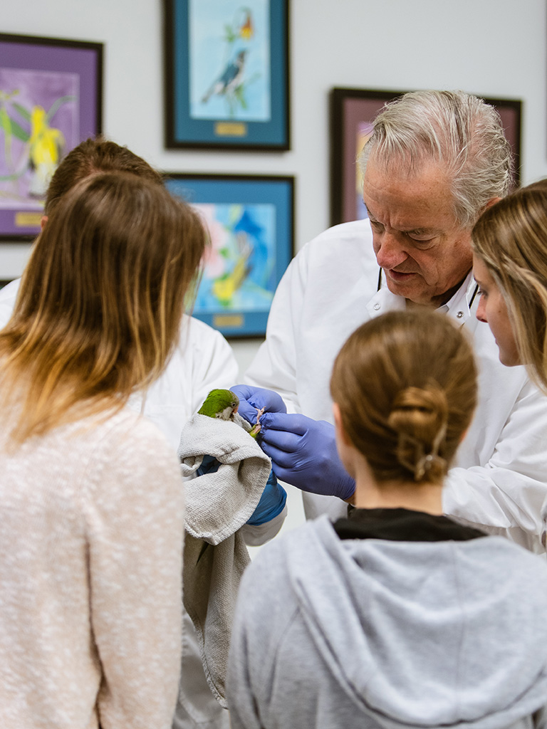
(from left to right) Dr. EV Voltura, Dr. Bianca Murphy (Harrison’s Bird Foods), Dr. Greg Harrison, Dr. Sarah Hamer, and Debra Turner (Senior Research Associate and Aviary Manager, Schubot Center). Photo by Alyssa Moore ’27, School of Veterinary Medicine & Biomedical Sciences
The Texas A&M Schubot Center for Avian Health has received a $250,000 donation from Harrison’s Bird Foods to advance avian health research, with a focus on nutrition, and allow the center to hire a postdoctoral researcher to help lead projects.
The new research to be conducted by the Schubot Center will determine how different diets contribute to the overall health of birds, including parrots. The planned work will advance not only the understanding of nutrition for caged and pet birds but also for bird populations in the wild.
The Schubot Center was founded in 1987 to improve the lives of birds and their environments through research, teaching, and outreach. It is housed within the Texas A&M School of Veterinary Medicine & Biomedical Sciences (VMBS) and has a membership of more than 85 faculty, staff, students, and clinicians across seven Texas A&M departments and three colleges/schools.
“We are extremely excited to be working with the professional staff at Schubot Center, the enthusiastic students, and with a group of long-term captive birds,” said Dr. Greg Harrison, the former first president of the Association of Avian Veterinarians and founder of Harrison’s Bird Foods. “Together, we have the opportunity to document the specific effects of diet in pet birds and be able to make scientific-based recommendations to the bird-owning public.”
“This new investment in nutritional research at the Schubot Center will allow us to take a comprehensive look at the health of birds exposed to different diets all in a controlled environment, including advanced bloodwork, microbiome analysis and even spectral analysis of feathers” said Dr. Sarah Hamer, the director and Richard Schubot Endowed Chair at the Schubot Center and a professor in both the VMBS’ Department of Veterinary Pathobiology (VTPB) and the Department of Veterinary Integrative Biosciences. “This donation will allow us to expand our center’s mission to improve bird health, and we are excited to engage veterinary students and undergraduates in all aspects of our center to provide them with unique training opportunities.”
As part of the donation, postdoctoral research associate Dr. EV Voltura will join the Schubot Center to help oversee part of the nutrition research.
Dr. Greg Harrison shows students how to conduct an avian wellness exam. Photo by Alyssa Moore ’27, School of Veterinary Medicine & Biomedical Sciences
“I’ve admired both Harrison’s Bird Foods and the work done at the Schubot Center for a long time,” Voltura said. “I knew that there was potential for an amazing research collaboration, and I was very fortunate to get to play a role in bringing everyone together.”
The donation will also fund additional nutrition studies focused on parrots led by Dr. Donald Brightsmith, also a VTPB professor and a member of the Schubot Center, in collaboration with Hill Country Aviaries and Harrison’s Bird Foods.
“We are excited to be working together to improve parrot diets,” Brightsmith said. “Many of the leading health concerns for captive parrots are related to diet, which is a similar issue in humans. Better diets means healthier parrots, so being able to combine the long history of nutritional products from Harrison’s Bird Foods with VBMS researchers should be a win for everyone. We are also excited about the prospect of working with Harrison’s on parrot conservation work and using their products to help rescued wild chicks and other parrots slated for release back into the wild.”
Members of Harrison’s Bird Foods visited the VMBS and the Schubot Center in mid-April to tour the center. Harrison gave an educational talk and demonstrated how to conduct avian wellness checks for Doctor of Veterinary Medicine students at the school.
“We were very happy to host Dr. Harrison and his team and are looking forward to continuing to promote avian nutrition and wellness together,” Hamer said.
###
For more information about the Texas A&M College of Veterinary Medicine & Biomedical Sciences, please visit our website at vetmed.tamu.edu or join us on Facebook, Instagram, and X.
Contact Information: Jennifer Gauntt, Director of VMBS Communications, Texas A&M College of Veterinary Medicine & Biomedical Sciences, jgauntt@cvm.tamu.edu,979-862-4216
The Texas A&M College of Veterinary Medicine & Biomedical Sciences (VMBS) recognized 71 students, faculty, and staff at the 2024 Honors & Awards Ceremony on April 24.
“The Texas A&M College of Veterinary Medicine & Biomedical Sciences would not be the great institution it is without the academic excellence and commitment of its faculty, staff, and students,” said Dr. John R. August, the Carl B. King Dean of Veterinary Medicine. “We applaud our recipients for their academic achievements and the impact that they are making through teaching, research, patient care, leadership, and service.”
Infographic by Aubree Herrold, School of Veterinary Medicine & Biomedical Sciences
Some of the most common reasons owners take their dogs to the vet have little or nothing to do with breed.
Many people assume that purebred dogs are more prone to disease than mixed-breed dogs, but a new study by the Dog Aging Project and led by researchers at the Texas A&M School of Veterinary Medicine and Biomedical Sciences (VMBS) and their Dog Aging Project collaborators across the country has busted this myth.
The study, published in the journal Frontiers In Veterinary Science, found that while certain dog breeds are prone to specific diseases, purebred and mixed-breed dogs are mostly equal when it comes to overall frequency of health condition diagnoses.
“There are several well-known diseases that frequently occur in specific dog breeds,” explained Dr. Kate Creevy, chief veterinary officer of the Dog Aging Project and a professor in the VMBS’ Department of Small Animal Clinical Sciences. “This has helped perpetuate the misconception that all purebred dogs are more prone to disease, but that is not the case.”
The study — which surveyed the owners of more than 27,000 companion dogs — also found that some of the most common diagnoses, like ear infections or osteoarthritis, occur in both purebreds and mixed-breed dogs.
Breaking Down The Data
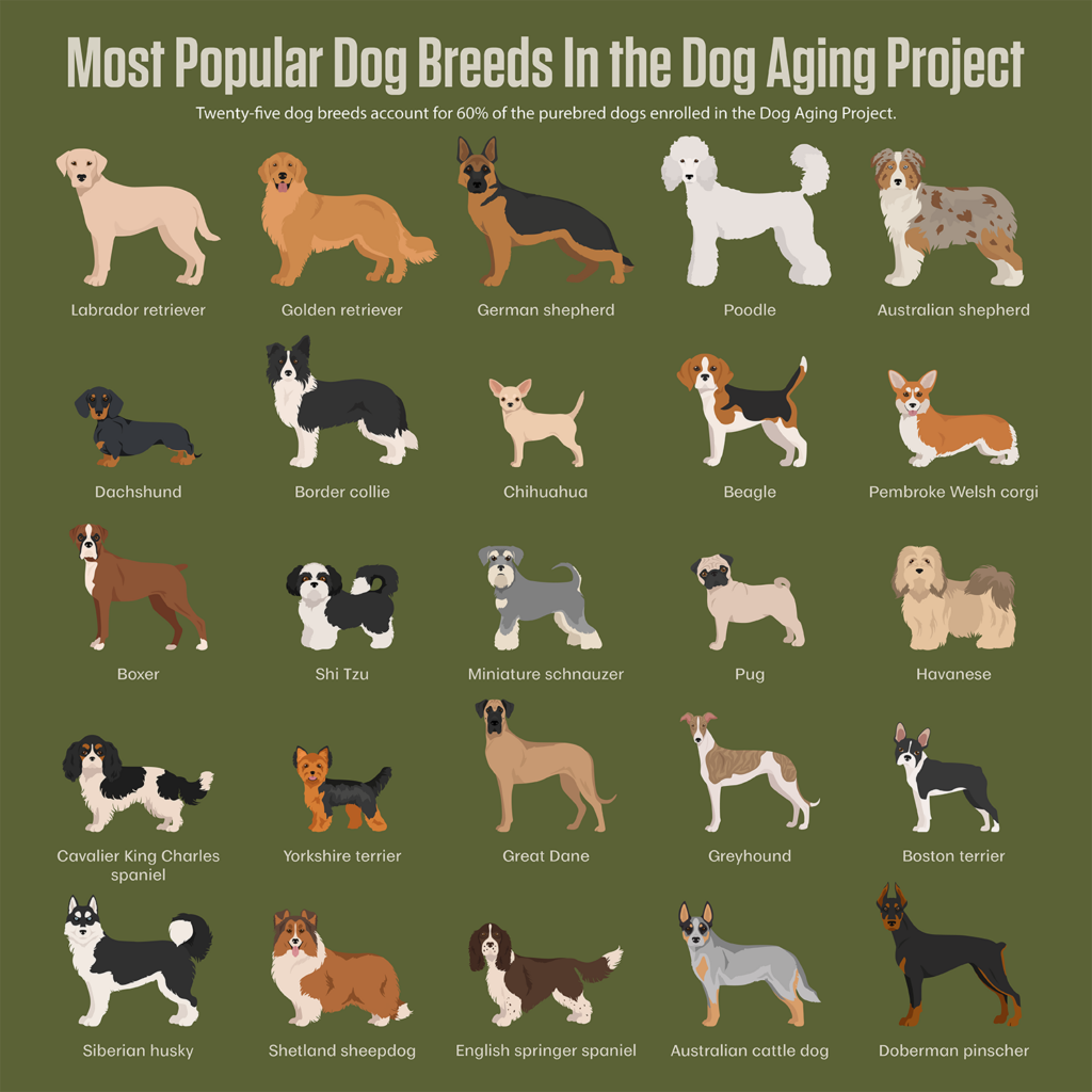
According to the study, 25 breeds make up about 60% of the purebred dog population within the Dog Aging Project. In order of popularity, those breeds are:
Labrador retriever
Golden retriever
German shepherd
Poodle
Australian shepherd
Dachshund
Border collie
Chihuahua
Beagle
Pembroke Welsh corgi
Boxer
Shi Tzu
Miniature schnauzer
Pug
Havanese
Cavalier King Charles spaniel
Yorkshire terrier
Great Dane
Greyhound
Boston terrier
Siberian husky
Shetland sheepdog
English springer spaniel
Australian cattle dog
Doberman pinscher
Within these 25 breeds, a total of 53 unique medical conditions make up the top owner-reported medical conditions.
“The medical conditions reported by owners of purebred dogs varied considerably,” Creevy said. “However, some conditions appeared frequently in the top 10 reported health conditions by breed.”
Across the 25 most popular breeds, those 10 conditions were:
Dental calculus (hardened plaque)
Dog bites
Extracted teeth
Giardia (a parasite)
Osteoarthritis
Seasonal allergies
Ear infection
Heart murmur
Fractured teeth
Cataracts
For mixed-breed dogs, the most common reported conditions were highly similar, with cataracts and heart murmur being replaced by torn/broken toenail and chocolate toxicity.
Some conditions, like dental calculus and osteoarthritis, appeared with roughly the same frequency in both purebred and mixed-breed dogs. Other conditions were more common in one than the other; extracted teeth and dog bites were more common in purebreds, versus ear infections in mixed-breed dogs.
“Out of the 53 medical conditions that owners reported, 26 did not differ significantly between mixed-breed and purebred dogs,” Creevy said.
Implications For Dog Owners
Dr. Kate Creevy
Ultimately, one of the most important findings from the study is that dog breed is only one aspect of pet health to consider when creating a pet’s care plan or researching what kind of dog to adopt.
“People should consider many factors when choosing a dog, including environment, lifestyle, social interactions and physical activity that will be available to the dog,” Creevy said. “Planning for both preventive veterinary care and medical care as the dog ages is also prudent. Dog owners should also talk with their primary care veterinarians about the kinds of medical problems to which their new dog might be particularly prone based on breed, size, sex, etc.”
As the study also showed, some of the most common reasons owners take their dogs to the vet have little or nothing to do with breed.
“Dental disease, allergies and osteoarthritis are among the most common conditions for all dogs,” Creevy said. “Owners should work with their primary care veterinarians on a plan to manage dental health. Regular exercise and maintaining lean body weight may help delay, prevent or lessen the impact of osteoarthritis.”
Expanding Dog Health Understanding
Though the study is already one of the largest cross-sectional studies of canine health, researchers at the Dog Aging Project are far from done examining its findings.
“We were surprised by the number of owners who reported that their dogs had experienced a bite from another dog,” Creevy said. “More investigation is needed to determine what this means and what particular factors might put an individual dog at risk.”
The DAP is a collaborative, community scientist-driven data-gathering research project that enrolls companion dogs from all backgrounds to study the effects of aging and gain a better understanding of what contributes to a long and healthy life for a dog.
The DAP continues to accept dogs of all breeds into the project. To date, more than 50,000 dogs have been enrolled.
Many of their research projects have led to translational studies that inform not only dog health, but also human health. To enroll your dog, or learn more, visit dogagingproject.org.
###
For more information about the Texas A&M College of Veterinary Medicine & Biomedical Sciences, please visit our website at vetmed.tamu.edu or join us on Facebook, Instagram, and X.
Contact Information: Jennifer Gauntt, Director of VMBS Communications, Texas A&M College of Veterinary Medicine & Biomedical Sciences, jgauntt@cvm.tamu.edu,979-862-4216
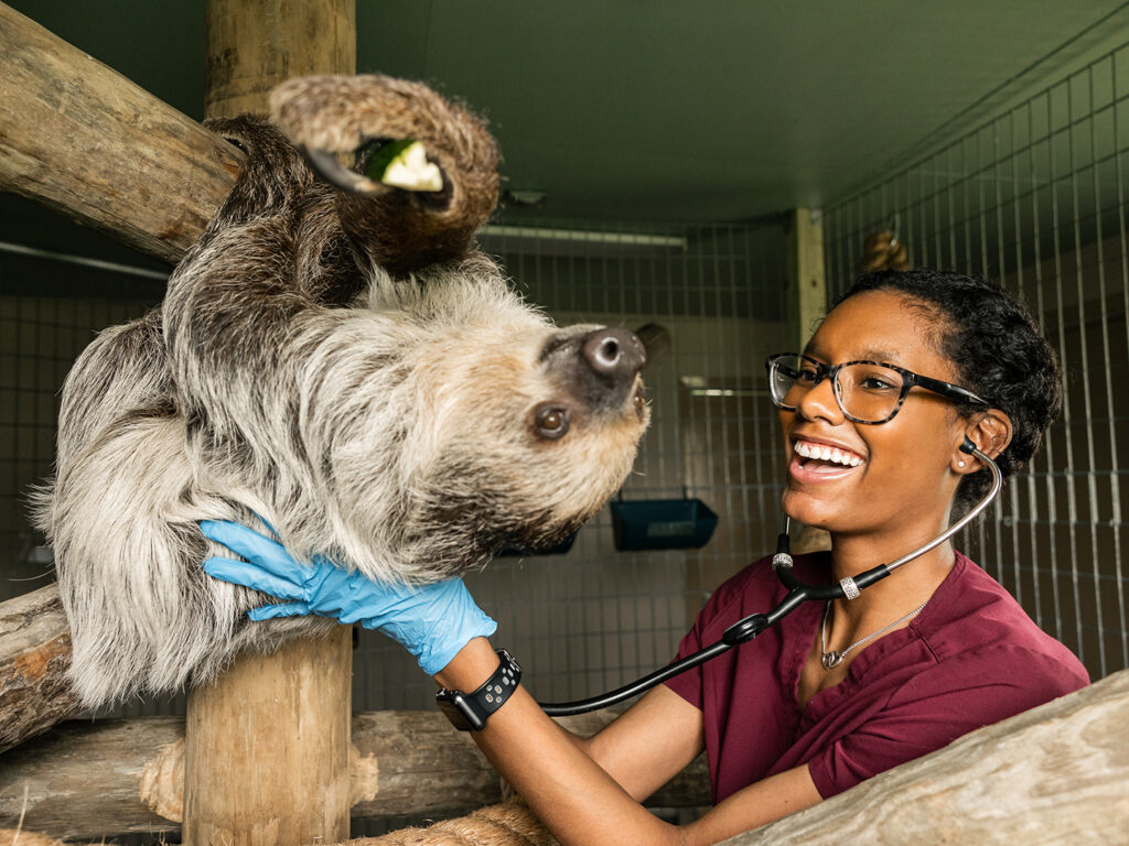
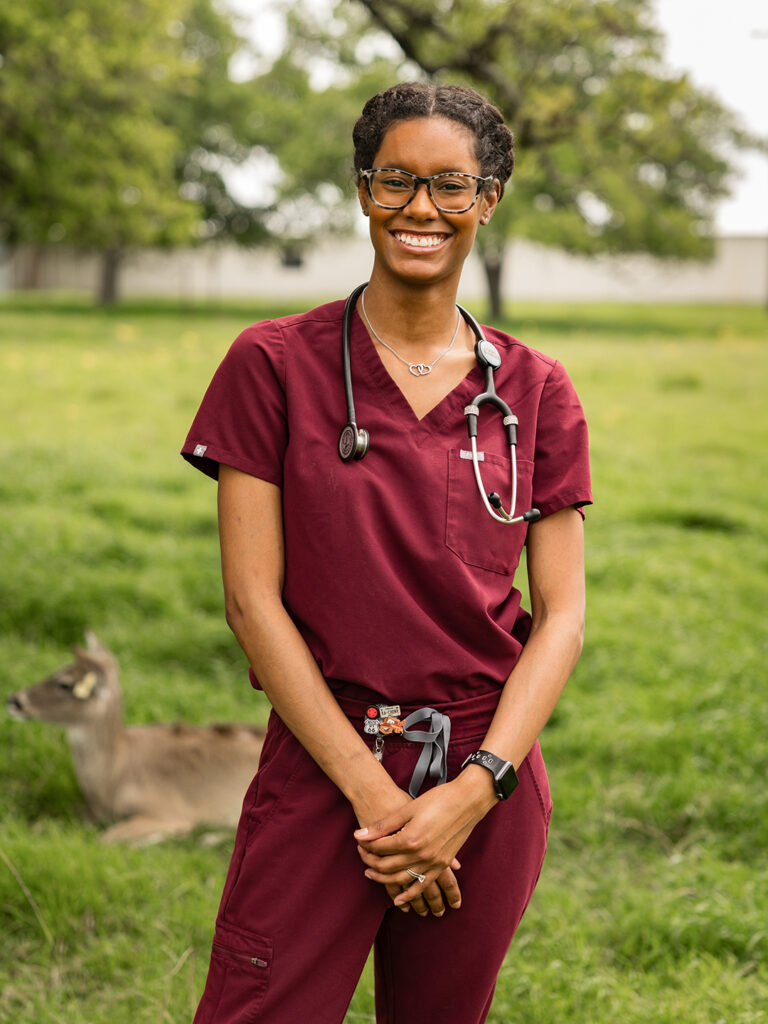
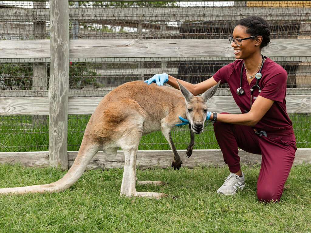
Amiri Fowler Cadena examines Michael Jr. Slow Poke Sloth at the Texas A&M Winnie Carter Wildlife Center. Photo by Jason Nitsch ’14, Texas A&M School of Veterinary Medicine & Biomedical Sciences
Over the course of four years as a Doctor of Veterinary Medicine (DVM) student at the Texas A&M School of Veterinary Medicine and Biomedical Sciences (VMBS), Amiri Fowler Cadena has been guided by a thirst for knowledge and desire to discover opportunities within the field of veterinary medicine.
While her veterinary journey has included ups and downs, Cadena has remained engaged — so much so that she now embodies a deep love for all aspects of animal care and will extend her love into the profession once she walks the graduation stage on May 8.
Embracing Versatility In Veterinary Medicine
From a young age, Cadena had a passion for animals, dreaming of a career as a zookeeper working with exotic species; however, a pivotal moment during her high school years changed the course of her ambitions.
“I was completely convinced that I was going to be a zookeeper, but then the high school I attended offered a veterinary program where students could shadow at clinics,” Cadena explained. “It was then that I discovered my passion for veterinary medicine.”
Through those shadowing experiences, Cadena discovered a new path within the field that resonated with her deeply.
“I remember always seeing cool cases, especially with the veterinary dentist,” Cadena said. “I saw her do dog braces, root canals and other general procedures, and I thought it was all amazing. At that point, I was particularly drawn to dentistry and surgery.”
Amiri Fowler Cadena Photo by Jason Nitsch ’14, Texas A&M School of Veterinary Medicine & Biomedical Sciences
After completing her undergraduate degree in biomedical sciences at Texas A&M University in 2019, Cadena took a gap year to work as a veterinary technician in a small animal clinic in Austin. This experience further fueled her passion for veterinary medicine and motivated her to explore more specialties within the field.
Upon being admitted into the VMBS’ Doctor of Veterinary Medicine (DVM) Class of 2024, Cadena eagerly participated in leadership roles and extracurricular activities, which not only enriched her experience but also shaped her into the confident and compassionate student she is today.
“I am very involved because I enjoy planning and being active in events,” Cadena said. “Coming into veterinary school, my passion for exotic animals led me to join the Zoo, Exotics, & Wildlife organization (ZEW) as a member my first year, working my way up to vice president my second year and president my third year. I am also the vice president for the Class of 2024, and I did a lot in helping plan class barbecues and Fur Balls (the annual DVM formal event) as well as setting up the mentorship program for incoming first-year students.”
Beyond academic and leadership demands, Cadena sought out additional extracurricular activities for both enjoyment and to more deeply explore different specialties within veterinary medicine, joining student organizations for dentistry, emergency and critical care (SVECCS), feline practitioners (SCAAFP), and surgery (SVSC).
“Before vet school, I was only into a small portion of what veterinary medicine can be, but now, with all of my new interests and experiences, I have been able to learn so many new things that I never understood or even knew were possible,” Cadena said. “I found that there are very few things in veterinary medicine that I don’t enjoy, so I came to a point where I realized I didn’t want to limit myself to just one specific area — I wanted to be able to do it all.”
Lessons Learned And Future Goals
Amiri Fowler Cadena examines a kangaroo at the Texas A&M Winnie Carter Wildlife Center. Photo by Jason Nitsch ’14, Texas A&M School of Veterinary Medicine & Biomedical Sciences
While she was enjoying her veterinary experience, navigating veterinary school wasn’t always easy for Cadena, so she sought support from various campus resources that helped her cope with the demands of the program.
“In my first year of vet school, I struggled to sleep because of stress and worry, but visiting our (on-site) counselors was one of the biggest resources that helped, especially since their services are free to A&M students,” Cadena explained. “I also sought help and advice from our veterinary faculty because they are all very nice and willing to talk to you. They are always willing to set you up with different opportunities that you would probably never think of or knew existed.”
Now, in her fourth year of veterinary school, Cadena made a deliberate decision to prioritize her clinical studies, taking a step back from some of her extracurricular commitments. Despite this adjustment, she continues to play a significant role as the vice president of the Class of 2024 and serves as a student member of the VMBS’ White Coats group, a team of veterinary student leaders who help with events such as orientation, the White Coat Ceremony and graduation.
“I have done better in veterinary school than I ever thought I would because I actually like what I’m doing and I enjoy the subjects,” Cadena said. “I feel incredibly fortunate to have chosen Texas A&M for my veterinary education.”
As her veterinary school journey draws to a close, Cadena prepares to join Town & Country Veterinary Hospital in Austin following commencement. The position will allow her to work with large animals, small animals and exotics, providing her with the flexibility to pursue her varied interests.
“Since coming to vet school, I have greatly expanded my knowledge of all species because the more I did, the more I realized that I love almost everything,” Cadena said. “Now, I’ll be able to share all that I have learned with my new clinic, broadening not only my own scope of practice but the scope of those around me as well.”
###
For more information about the Texas A&M College of Veterinary Medicine & Biomedical Sciences, please visit our website at vetmed.tamu.edu or join us on Facebook, Instagram, and X.
Contact Information: Jennifer Gauntt, Director of VMBS Communications, Texas A&M College of Veterinary Medicine & Biomedical Sciences, jgauntt@cvm.tamu.edu,979-862-4216